Review
. 2021 Jan;73(1):120-151.
doi: 10.1124/pharmrev.120.000082.

Post-Translational Modifications of G Protein-Coupled Receptors Control Cellular Signaling Dynamics in Space and Time

Affiliations
Review

Post-Translational Modifications of G Protein-Coupled Receptors Control Cellular Signaling Dynamics in Space and Time

Anand Patwardhan et al. Pharmacol Rev. 2021 Jan.

Abstract

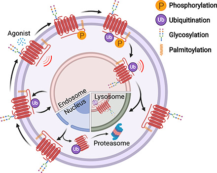
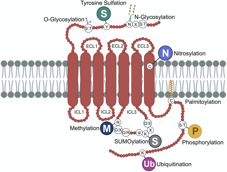
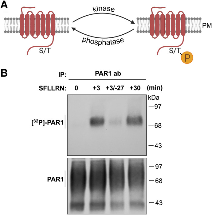
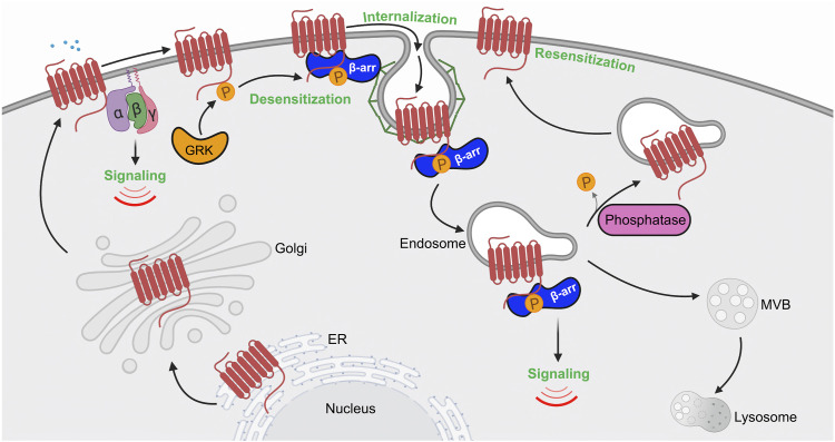
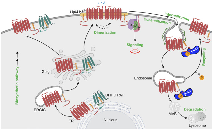
G protein-coupled receptors (GPCRs) are a large family comprising >800 signaling receptors that regulate numerous cellular and physiologic responses. GPCRs have been implicated in numerous diseases and represent the largest class of drug targets. Although advances in GPCR structure and pharmacology have improved drug discovery, the regulation of GPCR function by diverse post-translational modifications (PTMs) has received minimal attention. Over 200 PTMs are known to exist in mammalian cells, yet only a few have been reported for GPCRs. Early studies revealed phosphorylation as a major regulator of GPCR signaling, whereas later reports implicated a function for ubiquitination, glycosylation, and palmitoylation in GPCR biology. Although our knowledge of GPCR phosphorylation is extensive, our knowledge of the modifying enzymes, regulation, and function of other GPCR PTMs is limited. In this review we provide a comprehensive overview of GPCR post-translational modifications with a greater focus on new discoveries. We discuss the subcellular location and regulatory mechanisms that control post-translational modifications of GPCRs. The functional implications of newly discovered GPCR PTMs on receptor folding, biosynthesis, endocytic trafficking, dimerization, compartmentalized signaling, and biased signaling are also provided. Methods to detect and study GPCR PTMs as well as PTM crosstalk are further highlighted. Finally, we conclude with a discussion of the implications of GPCR PTMs in human disease and their importance for drug discovery. SIGNIFICANCE STATEMENT: Post-translational modification of G protein-coupled receptors (GPCRs) controls all aspects of receptor function; however, the detection and study of diverse types of GPCR modifications are limited. A thorough understanding of the role and mechanisms by which diverse post-translational modifications regulate GPCR signaling and trafficking is essential for understanding dysregulated mechanisms in disease and for improving and refining drug development for GPCRs.


Figures

None
Graphical abstract
Fig. 1.
Fig. 1.
GPCR post-translational modifications. GPCRs are seven-transmembrane proteins subjected to multiple types of PTMs on ECLs, ICLs, and the C-terminal domain. Here we show the most common sites of GPCR PTMs. PTMs that occur on ECLs include the following: N-glycosylation at asparagine (N)-X-serine (S)/threonine (T) sites, where X is any amino acid other than proline; O-glycosylation at S or T residues; and tyrosine (Y) sulfation. Nitrosylation has been shown to occur at transmembrane cysteine (C) residues. PTMs on intracellular loops include the following: SUMOylation on K residues present in the motif ψ-K-X-(D/E), where ψ is aliphatic amino acid, X is any amino acid, aspartic acid is D, and glutamic acid is E; methylation at arginine (R) residues of R-G-G or R-X-R sites, where glycine is G and X is any amino acid; and palmitoylation at cysteine (C). GPCR C-terminal PTMs include phosphorylation on S or T, rarely on Y residues, and ubiquitination (Ub) on specific K residues.
Fig. 2.
Fig. 2.
GPCR phosphorylation. (A) Agonist-activated GPCRs are phosphorylated at the cell surface primarily by GRKs, commonly at the C terminus on serine (S) or threonine (T) residues. Dephosphorylation of GPCRs is carried out by phosphatases. (B) Activation of PAR1 results in rapid phosphorylation as detected by immunoprecipitated (IP) [32]P-labeled PAR1 and autoradiography after 3 minutes of stimulation with peptide agonist 100 μM SFLLRN of PAR1 expressed in Rat1 fibroblasts. In PAR1-expressing cells stimulated with SFLLRN peptide for 3 minutes, followed by wash and chase for 27 minutes without agonist, PAR1 phosphorylation was no longer detectable, whereas continuous stimulation with SFLLRN for 30 minutes sustained phosphorylation. These findings suggest that PAR1 is subjected to phosphorylation and dephosphorylation. PAR1 protein from IPs detected by immunoblot with PAR1 antibody as shown in the bottom panel. ab, antibody; PM, plasma membrane; SFLLRN, Ser-Phe-Leu-Leu-Arg-Asn.
Fig. 3.
Fig. 3.
Model of GPCR regulation by phosphorylation. The schematic presents a classic view of GPCR regulation by phosphorylation in the cell. Agonist activation of a GPCR causes a conformational change that facilitates coupling to heterotrimeric G proteins (α, β, γ) and initiation of intracellular signaling cascades. Subsequently, GRKs are recruited and phosphorylate activated GPCRs at the C terminus, resulting in increased affinity and binding of β-arrestins (β-arr). β-arrestins compete with G protein binding to the same interhelical cavity localized within the cytoplasmic region of the GPCR. Once bound to the GPCR, β-arrestins prevent G protein coupling (desensitization) and facilitate association with clathrin and the endocytic machinery to promote internalization. Clathrin-coated pits bud inward and pinch off from the plasma membrane to form endocytic vesicles or endosomes. Once internalized, phosphorylation controls GPCRs’ capacity to nucleate the assembly of an endosomal β-arrestin signaling complex or, if dephosphorylated, GPCRs recycle from endosomes and return to the plasma membrane resulting in resensitization.
Fig. 4.
Fig. 4.
Ubiquitin modifying enzymes, ubiquitin linkages and detection. (A) Ubiquitination of substrate proteins is carried out sequentially by a ubiquitin (Ub)–activating enzyme E1, ∼38 ubiquitin-conjugating enzyme E2s, and >600 ubiquitin ligase E3 enzymes. Ubiquitin is enzymatically cleaved by ∼100 deubiquitinases to release ubiquitin back to cytosolic pool. (B) GPCRs are modified with different ubiquitin conjugations, including monoubiquitin (single or multiple monoubiquitin) and K48- or K63-linked polyubiquitin, which regulate distinct functions. Nonlysine ubiquitination has also been reported to occur on GPCRs. (C) Ubiquitination of endogenous PAR1 ubiquitination in endothelial cells after 7-minute stimulation with 10 nM thrombin (α-Th) detected by immunoblotting of immunoprecipitated (IP) PAR1 using anti-pan ubiquitin P4D1 antibody that detects multiple Ubn species. N-terminal proteolytic cleavage of PAR1 by thrombin results in reduced protein size of total protein detected by immunoblotting with PAR1 antibody (ab), bottom panel.
Fig. 5.
Fig. 5.
Model of GPCR regulation by ubiquitination. Biosynthesis and folding of GPCRs is monitored with stringent quality-control machinery in the ER, which targets misfolded GPCRs for ubiquitination and degradation through the ERAD-proteosomal pathway that releases ubiquitin (Ub) back to the cytosol. Properly folded GPCRs are delivered to the plasma membrane, where GPCRs are targeted for ubiquitination either basally or after agonist stimulation. Ubiquitination of GPCRs by E3 ligases has been implicated in agonist-induced internalization or basal receptor retention at the plasma membrane. Once internalized, ubiquitinated GPCR has multiple fates, including 1) recycling back to the plasma membrane, initiated by the action of deubiquitinases; 2) targeting for lysosomal degradation; and 3) ubiquitin-driven endosomal signaling.
Fig. 6.
Fig. 6.
GPCR maturation by glycosylation modifying enzymes and detection of glycosylation. (A) Glycans are covalently linked to GPCRs cotranslationally in the ER, mediate proper maturation, and facilitate expression at the plasma membrane (PM). N-glycans consist of N-acetylglucosamine (GlcNAc) attached to Asn (N) residues at the consensus Asn-X-Ser/Thr site, whereas O-glycosylation occurs at serine or threonine residues. Glycans are extensively trimmed in the Golgi and heterogeneous in nature. Misfolded GPCRs in the ER are cleared through endoplasmic-reticulum-associated protein degradation (ERAD)-proteasomal pathway. (B) Glycosylation of GPCRs occurs preferentially at the N-terminus and ECL2. (C) PAR1 expressed in HeLa cells treated with or without tunicamycin (Tunic), a global inhibitor of glycosylation, left panel. Mature PAR1 (PAR1M) migrates as multiple high-mobility bands, whereas treatment with tunicamycin results in a marked size shift of PAR1 to the predicted molecular weight, representative of unmodified or immature receptor (PAR1IM). PAR1 expressed in Rat1 fibroblasts treated with or without cycloheximide (CHX), a global inhibitor of protein synthesis, right panel. Mature PAR1M migrates predominantly as a high molecular weight species, with several lower migrating bands of partially modified or immature PAR1IM. Incubation with 100 μM SFLLRN agonist peptide for 2 hours results in mature PAR1M degradation but not PAR1IM. In non–SFLLRN-stimulated cells treated with CHX, immature PAR1IM is no longer detectable compared with mature PAR1M, which remains sensitive to SFLLRN-induced degradation. ab, antibody; endoH, endoglycosidase H; IP, immunoprecipitation; SFLLRN, Ser-Phe-Leu-Leu-Arg-Asn.
Fig. 7.
Fig. 7.
Model of GPCR regulation by glycosylation. GPCRs are extensively modified with N-glycosylation and O-glycosylation during biosynthesis and transport through the ER-Golgi en route to the plasma membrane. Glycosylation-deficient or misfolded GPCRs undergo ER-associated proteasomal degradation pathway. Glycosylation controls GPCR folding and maturation in the biosynthetic pathway, transport to the cell surface, signaling, ligand affinity, N-terminal cleavage, receptor dimerization, and internalization. Importantly, glycosylation controls GPCR biased signaling through direct modulation of the GPCR or in some cases the GPCR ligand. Glycosylation also regulates metalloprotease-mediated N-terminal cleavage of GPCR to influence biased signaling and modulates ligand-binding affinity by providing a larger and potentially more flexible binding surface of the GPCR. Dimerization of certain GPCRs is also positively modulated by glycosylation.
Fig. 8.
Fig. 8.
Palmitoylation modifying enzymes and detection of GPCR palmitoylation. (A) Palmitoyl-CoA, a derivative of palmitic acid, is a substrate of DHHC PATs, which catalyzes substrate palmitoylation through a two-step process, where a cysteine intermediate within a DHHC domain is autopalmitoylated; the palmitoyl moiety is then transferred to cysteine residues of the target protein. Palmitoylation is reversible. APTs remove the palmitoylation moiety from substrate proteins. (B) HeLa cells expressing PAR1 wild type (WT) and mutant, in which cysteine (C)387 and C388 were converted to alanine (A), were metabolically labeled with [3H]-palmitate and either left untreated or treated with 100 μM SFLLRN peptide agonist for various times. Cells were lysed, PAR1 was immunoprecipitated (IP) and subjected to autoradiography to visualize [3H]palmitate-labeled PAR1 or immunoblot (IB) to detect total PAR1 protein with PAR1 antibody (ab). (C) A DHHC PATs are located at the ER, Golgi, and plasma membrane. The juxtaposition of the PAT to the target GPCR facilitates palmitoylation. GPCR palmitoylation on C-terminal tail cysteines embeds a region in the membrane creating a fourth intracellular loop and, in some cases, facilitates GPCR localization to lipid rafts. Depalmitoylation of substrate proteins including GPCRs is mediated by APT, which itself may be subjected to palmitoylation to facilitate membrane localization. SFLLRN, Ser-Phe-Leu-Leu-Arg-Asn.
Fig. 9.
Fig. 9.
Model of GPCR regulation by palmitoylation. GPCRs are palmitoylated during biosynthesis and can occur at the endoplasmic reticulum (ER), endoplasmic-reticulum–Golgi intermediate compartment (ERGIC), Golgi and the plasma membrane, where DHHC PATs are known to be localized. GPCR palmitoylation regulates partitioning into membrane microdomains enriched in cholesterol such as lipid rafts and caveolae. GPCR palmitoylation has also been implicated in receptor dimerization as well as G protein coupling. Palmitoylation of GPCRs can further influence β-arrestin (β-arr) recruitment and receptor internalization. GPCR palmitoylation is also important for regulating receptor recycling and thereby prevents lysosomal degradation.

Similar articles

Cited by

References

    1. Adachi N, Hess DT, McLaughlin P, Stamler JS. (2016) S-palmitoylation of a novel site in the β2-adrenergic receptor associated with a novel intracellular itinerary. J Biol Chem 291:20232–20246. - PMC - PubMed
    1. Adams MN, Christensen ME, He Y, Waterhouse NJ, Hooper JD. (2011) The role of palmitoylation in signalling, cellular trafficking and plasma membrane localization of protease-activated receptor-2. PLoS One 6:e28018. - PMC - PubMed
    1. Alvarez CE. (2008) On the origins of arrestin and rhodopsin. BMC Evol Biol 8:222. - PMC - PubMed
    1. Alvaro CG, O’Donnell AF, Prosser DC, Augustine AA, Goldman A, Brodsky JL, Cyert MS, Wendland B, Thorner J. (2014) Specific α-arrestins negatively regulate Saccharomyces cerevisiae pheromone response by down-modulating the G-protein-coupled receptor Ste2. Mol Cell Biol 34:2660–2681. - PMC - PubMed
    1. Arakaki AKS, Pan WA, Lin H, Trejo J. (2018a) The α-arrestin ARRDC3 suppresses breast carcinoma invasion by regulating G protein-coupled receptor lysosomal sorting and signaling. J Biol Chem 293:3350–3362. - PMC - PubMed

Publication types

Substances