Review
. 2021 Feb;31(2):107-125.
doi: 10.1038/s41422-020-00441-1. Epub 2020 Dec 2.

Ferroptosis: molecular mechanisms and health implications

Affiliations
Review

Ferroptosis: molecular mechanisms and health implications

Daolin Tang et al. Cell Res. 2021 Feb.

Abstract

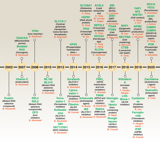
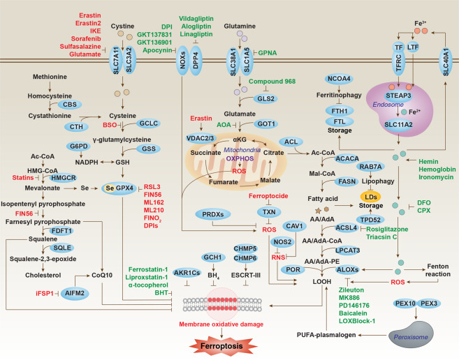
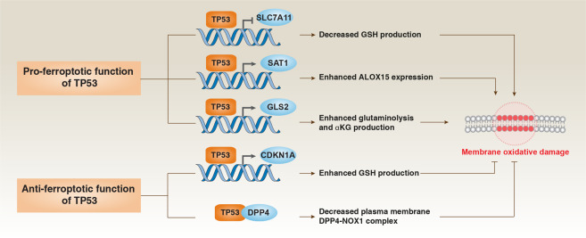
Cell death can be executed through different subroutines. Since the description of ferroptosis as an iron-dependent form of non-apoptotic cell death in 2012, there has been mounting interest in the process and function of ferroptosis. Ferroptosis can occur through two major pathways, the extrinsic or transporter-dependent pathway and the intrinsic or enzyme-regulated pathway. Ferroptosis is caused by a redox imbalance between the production of oxidants and antioxidants, which is driven by the abnormal expression and activity of multiple redox-active enzymes that produce or detoxify free radicals and lipid oxidation products. Accordingly, ferroptosis is precisely regulated at multiple levels, including epigenetic, transcriptional, posttranscriptional and posttranslational layers. The transcription factor NFE2L2 plays a central role in upregulating anti-ferroptotic defense, whereas selective autophagy may promote ferroptotic death. Here, we review current knowledge on the integrated molecular machinery of ferroptosis and describe how dysregulated ferroptosis is involved in cancer, neurodegeneration, tissue injury, inflammation, and infection.


Conflict of interest statement

The authors declare no competing interests.

Figures

Fig. 1
Fig. 1. Timeline of ferroptosis research.
A brief history of molecular and pharmacological modulators of ferroptosis.
Fig. 2
Fig. 2. Core molecular machinery and signaling regulation of ferroptosis.
Ferroptosis can occur through two major pathways, the extrinsic or transporter-dependent pathway (e.g., decreased cysteine or glutamine uptake and increased iron uptake), and the intrinsic or enzyme-regulated pathway (e.g., the inhibition of GPX4).
Fig. 3
Fig. 3. Immune features of ferroptosis.
Ferroptotic cell death can lead to different immune and inflammatory reactions through the release and activation of damage-associated molecular patterns (e.g., HMGB1 and DNA) or lipid oxidation products (e.g., 4HNE, oxPLs, LTB4, LTC4, LTD4, and PGE2) in immune cells (e.g., macrophages, monocytes, and neutrophils) via different intracellular signal transduction pathways.
Fig. 4
Fig. 4. Role of autophagy in ferroptosis.
a The mechanism of non-selective macroautophagy/autophagy induced by ferroptosis activator (e.g., erastin and RSL3). b, c Certain selective types of autophagy (e.g., ferritinophagy, lipophagy, clockophagy and mitophagy) (b) and chaperone-mediated autophagy (c) promote oxidative damage-dependent ferroptosis through the degradation of ferroptosis repressors (e.g., ferritin, ARNTL/BMAL1, lipid droplets and GPX4).
Fig. 5
Fig. 5. NFE2L2 in ferroptosis.
a Under normal conditions, a low level of NFE2L2 is primarily maintained by KEAP1-mediated proteasomal degradation. b Following ferroptosis stress, the NFE2L2 protein is stabilized and initiates a multi-step activation pathway, including nuclear translocation, heterodimerization with its partner MAF protein, recruitment of transcription coactivators and subsequent binding to the antioxidant response element (ARE) of the target gene promoter. SQSTM1 can stabilize NFE2L2 by inactivating KEAP1 through autophagic degradation.
Fig. 6
Fig. 6. TP53 in ferroptosis.
TP53 plays a context-dependent role in the regulation of membrane oxidative damage in ferroptosis. On the one hand, TP53 can enhance ferroptosis by inhibiting SLC7A11 expression or promoting SAT1 and GLS2 expression. On the other hand, TP53 can inhibit ferroptosis by inhibiting DPP4 activity or inducing CDKN1A expression.
Fig. 7
Fig. 7. Dual role of ferroptosis in tumor immunity.
a CD8+ T cell-mediated IFNG release inhibits SLC7A11 expression in cancer cells through activation of the STAT1 pathway, thereby inducing tumor cell ferroptosis. b Ferroptotic cancer cell-mediated KRASG12D release increases M2 macrophage polarization through activation of the STAT3 pathway, thereby limiting antitumor immunity.

References

    1. Galluzzi L, et al. Molecular mechanisms of cell death: recommendations of the Nomenclature Committee on Cell Death 2018. Cell Death Differ. 2018;25:486–541. - PMC - PubMed
    1. Tang D, Kang R, Berghe TV, Vandenabeele P, Kroemer G. The molecular machinery of regulated cell death. Cell Res. 2019;29:347–364. - PMC - PubMed
    1. Stockwell BR, et al. Ferroptosis: a regulated cell death nexus linking metabolism, redox biology, and disease. Cell. 2017;171:273–285. - PMC - PubMed
    1. Bieri JG. An effect of selenium and cystine on lipide peroxidation in tissues deficient in vitamin E. Nature. 1959;184:1148–1149. - PubMed
    1. Moore B, Hawkes JL. An investigation of the toxic actions of dilute solutions of the salts of certain heavy metals (viz.: copper, iron, nickel, cobalt, manganese, zinc, silver, and lead) upon the Bacillus Typhosus, with a view to practical application in the purification of shell-fish. Biochem. J. 1908;3:313–345. - PMC - PubMed

Publication types