Case series of three patients with hereditary diffuse gastric cancer in a single family: Three case reports and review of literature
- PMID: 33268956
- PMCID: PMC7673959
- DOI: 10.3748/wjg.v26.i42.6689
Case series of three patients with hereditary diffuse gastric cancer in a single family: Three case reports and review of literature
Abstract
Background: Hereditary diffuse gastric cancer (HDGC) is a familial cancer syndrome often associated with germline mutations in the CDH1 gene. However, the frequency of CDH1 mutations is low in patients with HDGC in East Asian countries. Herein, we report three cases of HDGC harboring a missense CDH1 variant, c.1679C>G, from a single Japanese family.
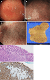
Case summary: A 26-year-old female (Case 1) and a 51-year-old male (father of Case 1), who had a strong family history of gastric cancer, were diagnosed with advanced diffuse gastric cancer. After genetic counselling, a 25-year-old younger brother of Case 1 underwent surveillance esophagogastroduodenoscopy that detected small signet ring cell carcinoma foci as multiple pale lesions in the gastric mucosa. Genetic analysis revealed a CDH1 c.1679C>G variant in all three patients.
Conclusion: It is important for individuals suspected of having HDGC to be actively offered genetics evaluation. This report will contribute to an increased awareness of HDGC.
Keywords: CDH1; Case report; E-cadherin; Endoscopic findings; Hereditary diffuse gastric cancer; Signet ring cell carcinoma.
©The Author(s) 2020. Published by Baishideng Publishing Group Inc. All rights reserved.
Conflict of interest statement
Conflict-of-interest statement: The authors have no conflicts to declare.
Figures


References
-
- Ferlay J, Colombet M, Soerjomataram I, Mathers C, Parkin DM, Piñeros M, Znaor A, Bray F. Estimating the global cancer incidence and mortality in 2018: GLOBOCAN sources and methods. Int J Cancer. 2019;144:1941–1953. - PubMed
-
- van der Post RS, Vogelaar IP, Carneiro F, Guilford P, Huntsman D, Hoogerbrugge N, Caldas C, Schreiber KE, Hardwick RH, Ausems MG, Bardram L, Benusiglio PR, Bisseling TM, Blair V, Bleiker E, Boussioutas A, Cats A, Coit D, DeGregorio L, Figueiredo J, Ford JM, Heijkoop E, Hermens R, Humar B, Kaurah P, Keller G, Lai J, Ligtenberg MJ, O'Donovan M, Oliveira C, Pinheiro H, Ragunath K, Rasenberg E, Richardson S, Roviello F, Schackert H, Seruca R, Taylor A, Ter Huurne A, Tischkowitz M, Joe ST, van Dijck B, van Grieken NC, van Hillegersberg R, van Sandick JW, Vehof R, van Krieken JH, Fitzgerald RC. Hereditary diffuse gastric cancer: updated clinical guidelines with an emphasis on germline CDH1 mutation carriers. J Med Genet. 2015;52:361–374. - PMC - PubMed
-
- Guilford P, Hopkins J, Harraway J, McLeod M, McLeod N, Harawira P, Taite H, Scoular R, Miller A, Reeve AE. E-cadherin germline mutations in familial gastric cancer. Nature. 1998;392:402–405. - PubMed
-
- Wang Y, Song JP, Ikeda M, Shinmura K, Yokota J, Sugimura H. Ile-Leu substitution (I415L) in germline E-cadherin gene (CDH1) in Japanese familial gastric cancer. Jpn J Clin Oncol. 2003;33:17–20. - PubMed
Publication types
MeSH terms
Substances
LinkOut - more resources
Full Text Sources
Medical
Miscellaneous

