

Multilocular Thymic Cyst in a Young, Otherwise Healthy Woman: A Case Report
- PMID: 33269141
- PMCID: PMC7704160
- DOI: 10.7759/cureus.11210
Multilocular Thymic Cyst in a Young, Otherwise Healthy Woman: A Case Report
Abstract
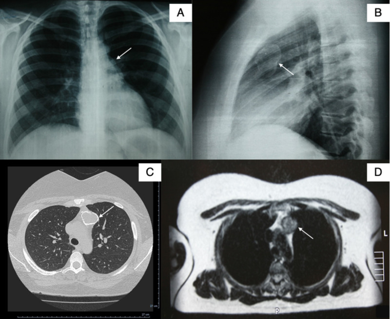
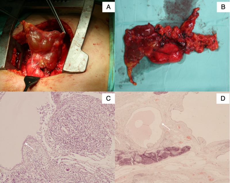
Thymic cysts are rare lesions, accounting approximately for 1% of all mediastinal masses. We report a case of a 36-year old woman who presented preoperatively with a calcified mass shadow found on a routine chest radiograph X-ray. After further investigation with chest computed tomography (CT), magnetic resonance imaging (MRI), and tests for Myasthenia gravis, a benign mediastinal cyst was diagnosed and the patient underwent median sternotomy and complete surgical excision of the lesion. The histological examination described a multilocular thymic cyst. Thymic cysts are usually associated with thymic epithelial tumors, such as thymomas, or multisystemic morbid conditions such as human immunodeficiency virus (HIV) infection, rheumatologic disease, and Myasthenia gravis. At all follow-up examinations to date, the patient remains healthy.
Keywords: cyst; mediastinal; multilocular; thymic.
Copyright © 2020, Damaskos et al.
Conflict of interest statement
The authors have declared that no competing interests exist.
Figures
References
-
- Clinical spectrum of mediastinal cysts. Takeda S, Miyoshi S, Minami M, Mitsunori O, Akira M, Hikaru M. Chest. 203;124:125–132. - PubMed
-
- Multilocular thymic cyst: an acquired reactive process. Study of 18 cases. Suster S, Rosai J. https://pubmed.ncbi.nlm.nih.gov/2006719. Am J Surg Pathol. 1991;15:388–398. - PubMed
-
- Asymptomatic giant thymic cyst treated with VATS. Çelik B, Gürz S, Elmalı M, Süllü Y. Curr Thorac Surg. 2017;2:67–69.
-
- Multilocular mediastinal cyst with rim calcification: report of a case. Osaki T, Nakagawa M. Surg Today. 2008;38:52–55. - PubMed
-
- Mediastinal cyst with rim calcification. Sugimoto S, Misao T, Nakano H, Yamane M. Jpn J Thorac Cardiovasc Surg. 2004;52:261–263. - PubMed
Publication types
LinkOut - more resources
Full Text Sources