. 2020 Nov 25;6(11):2046-2052.
doi: 10.1021/acscentsci.0c00855. Epub 2020 Sep 23.

The SARS-COV-2 Spike Protein Binds Sialic Acids and Enables Rapid Detection in a Lateral Flow Point of Care Diagnostic Device

Affiliations

The SARS-COV-2 Spike Protein Binds Sialic Acids and Enables Rapid Detection in a Lateral Flow Point of Care Diagnostic Device

Alexander N Baker et al. ACS Cent Sci. .

Erratum in

Abstract

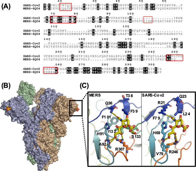
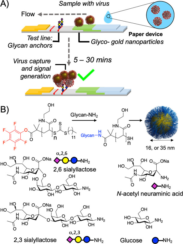
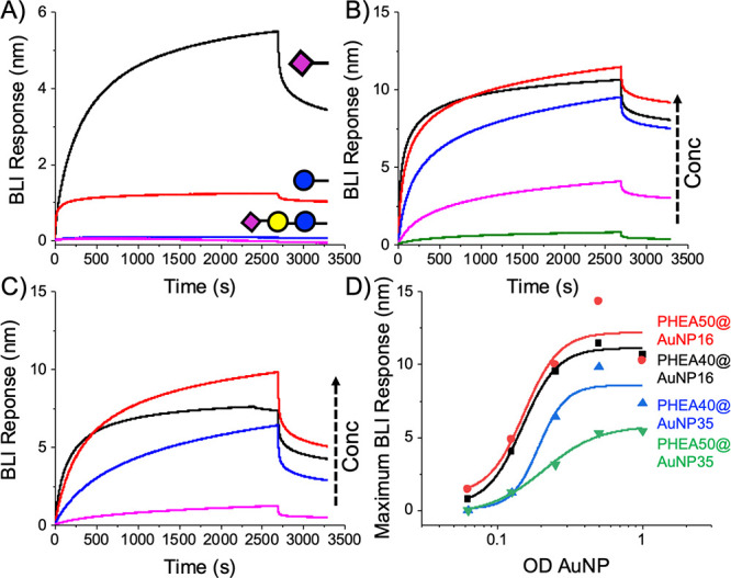
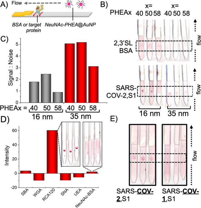
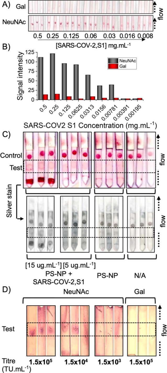
There is an urgent need to understand the behavior of the novel coronavirus (SARS-COV-2), which is the causative agent of COVID-19, and to develop point-of-care diagnostics. Here, a glyconanoparticle platform is used to discover that N-acetyl neuraminic acid has affinity toward the SARS-COV-2 spike glycoprotein, demonstrating its glycan-binding function. Optimization of the particle size and coating enabled detection of the spike glycoprotein in lateral flow and showed selectivity over the SARS-COV-1 spike protein. Using a virus-like particle and a pseudotyped lentivirus model, paper-based lateral flow detection was demonstrated in under 30 min, showing the potential of this system as a low-cost detection platform.


Conflict of interest statement

The authors declare the following competing financial interest(s): M.I.G., A.N.B., and S.J.R. are named inventors of a patent relating to this work. R.F. is a shareholder in Iceni diagnostics who part-funded this work.

Figures

Figure 1
Figure 1
(A) Sequence alignment between the S1 domains of the SARS-COV-2 and MERS spike proteins. Regions important for sialic acid binding are highlighted by red boxes; (B) Model showing the hypothesized sialic acid binding sites (yellow CPK coloring) for the SARS-COV-2 spike protein trimer; (C) A comparison between the sialic acid binding sites from MERS (PDB entry 6Q04) and the SARS-COV-2 model (PDB entry 6VSB) in complex with a2,3′-sialyllactose.
Figure 2
Figure 2
Design concept for glyco-lateral flow devices. (A) Lateral flow assay for virus, using glycan capture units; (B) Synthetic procedure for glyconanoparticles.
Figure 3
Figure 3
Biolayer interferometry analysis of SARS-COV-2 spike protein with glyconanoparticles. (A) Screening using PHEA50@AuNP35 at OD = 1; Dose-dependent binding of NeuNAc-PHEA50 using (B) @AuNP16 and (C) @AuNP35. OD = 1 (−), 0.5(-), 0.25(-), 0.125(-); (D) Binding curves.
Figure 4
Figure 4
Half lateral flow analysis of NeuNAcPHEAx@AuNPy particles. (A) Half lateral flow assay setup with target protein immobilized on the test line; (B) Effect of polymer chain length and particle size on lateral flow binding; (C) Signal:noise analysis; (D) Selectivity of NeuNAcPHEA50@AuNP35 against a panel of lectins (inset example LFD strips). (E) Selectivity of NeuNAcPHEA50@AuNP35 against S1 protein from different coronavirus strains. Data is the mean from 3 repeats. Original LFD strips are in the SI. Test lines are within the dashed-line box. 2,3′SL-BSA = 2,3′-sialyllactose-functionalized BSA.
Figure 5
Figure 5
(A) Detection limit analysis of galactose or NeuNAc functionalized AuNPs against immobilized SARS-COV-2,S1 using “half LFD” assays; (B) Signal intensity analysis; (C) Dipstick lateral flow tests using NeuNAcPHEA50@AuNP35 and BSA-NeuNAc as the test line and RCA120 as the control line. PS-NP = 100 nm polystyrene colloid, or + SARS-COV-2,S1. N/A is with no polystyrene analyte. (D) Half lateral flow analysis of SARS-COV-2 Spike pseudotyped lentivirus against NeuNAcPHEA50@AuNP35 or GalPHEA50@AuNP35 nanoparticles. In each image, the test line region is indicated by the dashed box. Complete original images are in Supporting Information.

Similar articles

Cited by

References

    1. Zhou P.; Yang X.-L.; Wang X.-G.; Hu B.; Zhang L.; Zhang W.; Si H.-R.; Zhu Y.; Li B.; Huang C.-L.; Chen H.-D.; Chen J.; Luo Y.; Guo H.; Jiang R.-D.; Liu M.-Q.; Chen Y.; Shen X.-R.; Wang X.; Zheng X.-S.; Zhao K.; Chen Q.-J.; Deng F.; Liu L.-L.; Yan B.; Zhan F.-X.; Wang Y.-Y.; Xiao G.-F.; Shi Z.-L. A Pneumonia Outbreak Associated with a New Coronavirus of Probable Bat Origin. Nature 2020, 579 (7798), 270–273. 10.1038/s41586-020-2012-7. - DOI - PMC - PubMed
    1. Xie X.; Zhong Z.; Zhao W.; Zheng C.; Wang F.; Liu J. Ches CT for Typical 2019-NCoV Pneumonia: Relationship to Negative RT-PCR Testing. Radiology 2020, 296, E41.10.1148/radiol.2020200343. - DOI - PMC - PubMed
    1. Li Y.; Yao L.; Li J.; Chen L.; Song Y.; Cai Z.; Yang C. Stability Issues of RT-PCR Testing of SARS-CoV-2 for Hospitalized Patients Clinically Diagnosed with COVID-19. J. Med. Virol. 2020, 92, 903.10.1002/jmv.25786. - DOI - PMC - PubMed
    1. Huang P.; Liu T.; Huang L.; Liu H.; Lei M.; Xu W.; Hu X.; Chen J.; Liu B. Use of Chest CT in Combination with Negative RT-PCR Assay for the 2019 Novel Coronavirus but High Clinical Suspicion. Radiology 2020, 295, 22–23. 10.1148/radiol.2020200330. - DOI - PMC - PubMed
    1. Hase R.; Kurita T.; Muranaka E.; Sasazawa H.; Mito H.; Yano Y. A Case of Imported COVID-19 Diagnosed by PCR-Positive Lower Respiratory Specimen but with PCR-Negative Throat Swabs. Infect. Dis. (Auckl). 2020, 52 (6), 423–426. 10.1080/23744235.2020.1744711. - DOI - PMC - PubMed