[Preprint]. 2020 Nov 24:2020.11.24.393405.
doi: 10.1101/2020.11.24.393405.

Fragment Binding to the Nsp3 Macrodomain of SARS-CoV-2 Identified Through Crystallographic Screening and Computational Docking

Affiliations

Fragment Binding to the Nsp3 Macrodomain of SARS-CoV-2 Identified Through Crystallographic Screening and Computational Docking

Marion Schuller et al. bioRxiv. .

Update in

  • Fragment binding to the Nsp3 macrodomain of SARS-CoV-2 identified through crystallographic screening and computational docking.
    Schuller M, Correy GJ, Gahbauer S, Fearon D, Wu T, Díaz RE, Young ID, Carvalho Martins L, Smith DH, Schulze-Gahmen U, Owens TW, Deshpande I, Merz GE, Thwin AC, Biel JT, Peters JK, Moritz M, Herrera N, Kratochvil HT; QCRG Structural Biology Consortium; Aimon A, Bennett JM, Brandao Neto J, Cohen AE, Dias A, Douangamath A, Dunnett L, Fedorov O, Ferla MP, Fuchs MR, Gorrie-Stone TJ, Holton JM, Johnson MG, Krojer T, Meigs G, Powell AJ, Rack JGM, Rangel VL, Russi S, Skyner RE, Smith CA, Soares AS, Wierman JL, Zhu K, O'Brien P, Jura N, Ashworth A, Irwin JJ, Thompson MC, Gestwicki JE, von Delft F, Shoichet BK, Fraser JS, Ahel I. Schuller M, et al. Sci Adv. 2021 Apr 14;7(16):eabf8711. doi: 10.1126/sciadv.abf8711. Print 2021 Apr. Sci Adv. 2021. PMID: 33853786 Free PMC article.

Abstract

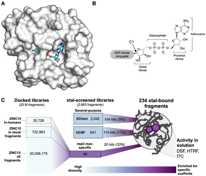
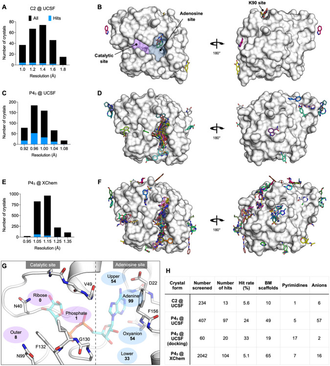
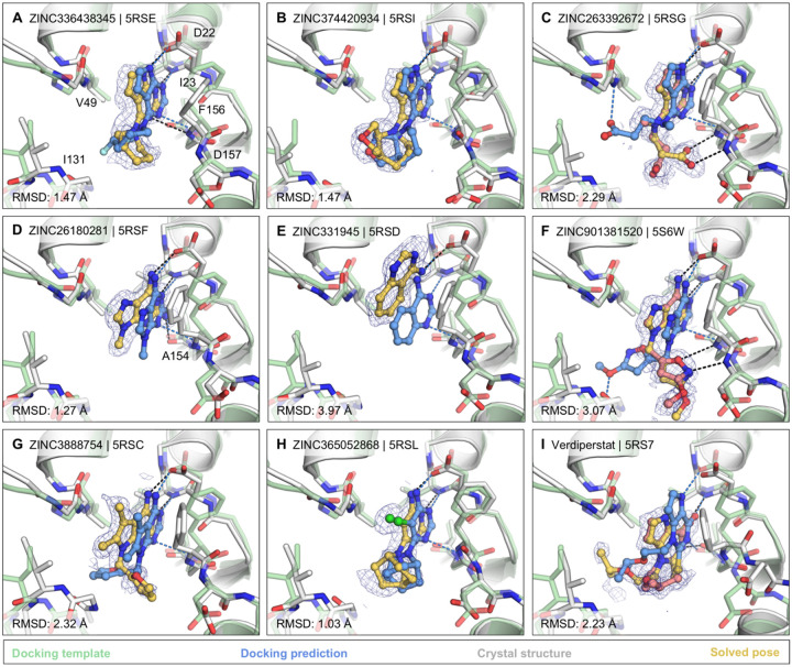
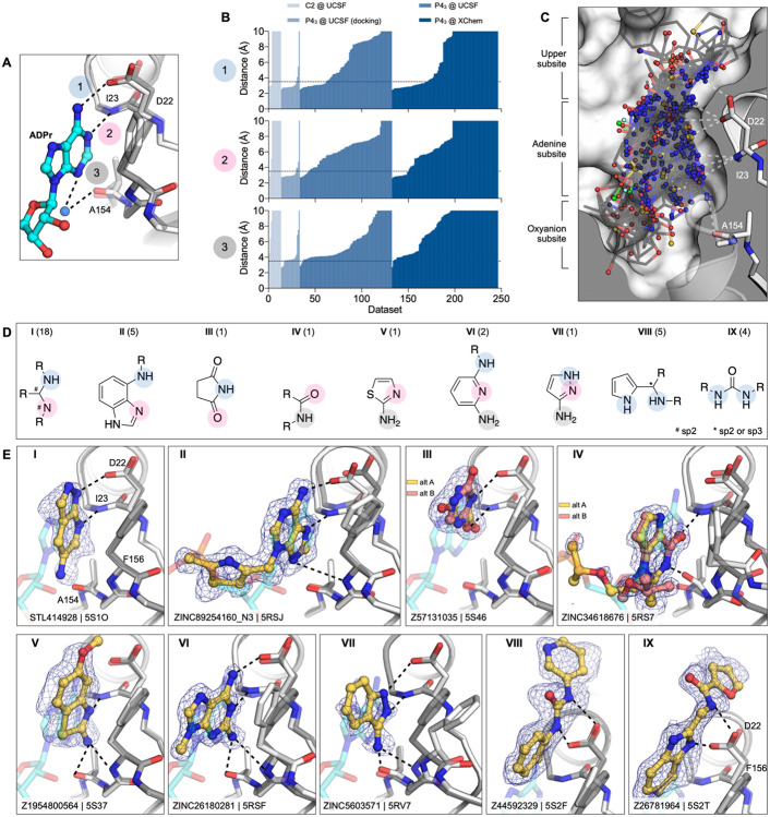
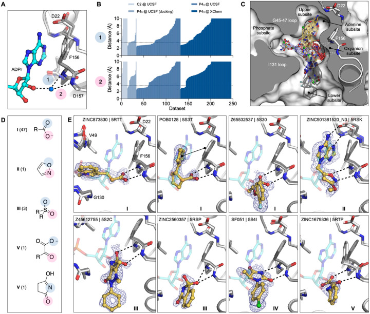
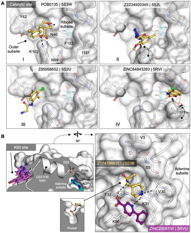
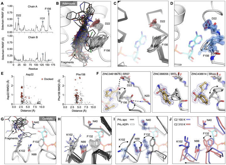
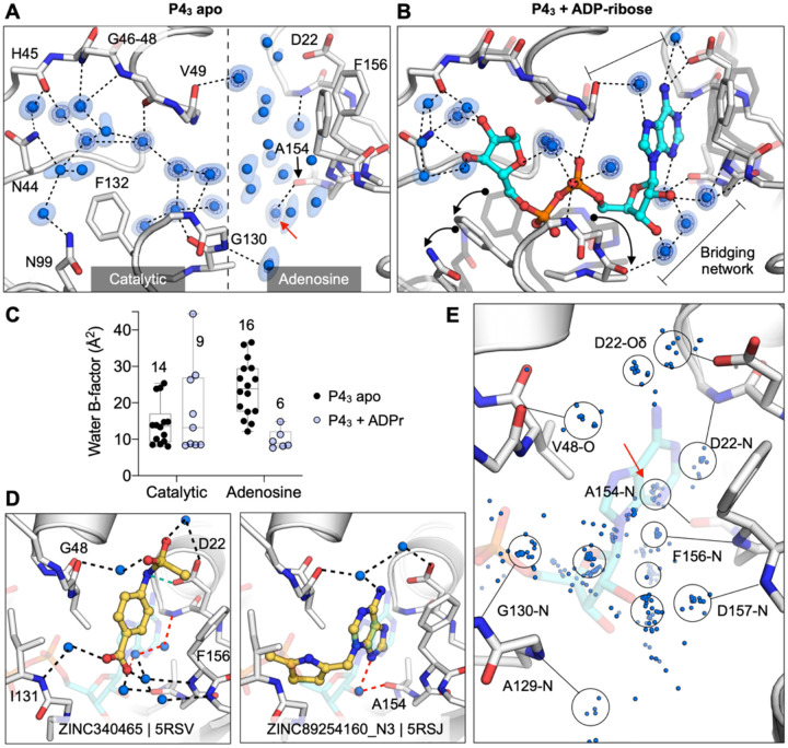
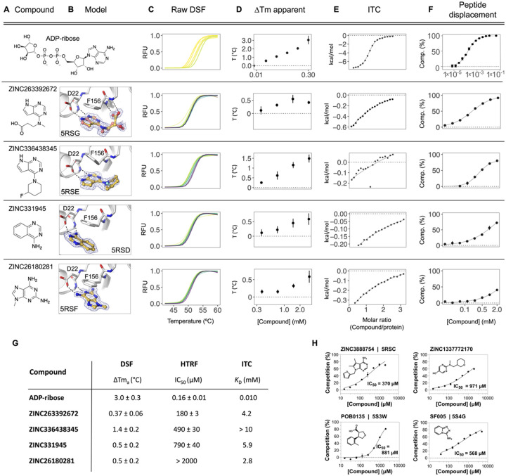
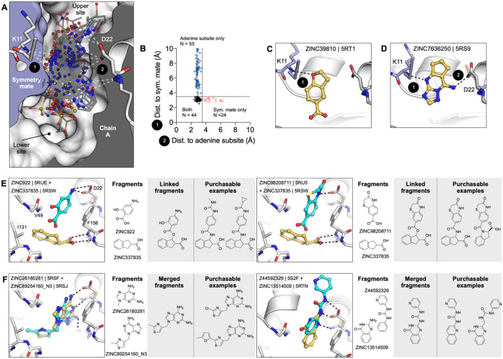
The SARS-CoV-2 macrodomain (Mac1) within the non-structural protein 3 (Nsp3) counteracts host-mediated antiviral ADP-ribosylation signalling. This enzyme is a promising antiviral target because catalytic mutations render viruses non-pathogenic. Here, we report a massive crystallographic screening and computational docking effort, identifying new chemical matter primarily targeting the active site of the macrodomain. Crystallographic screening of diverse fragment libraries resulted in 214 unique macrodomain-binding fragments, out of 2,683 screened. An additional 60 molecules were selected from docking over 20 million fragments, of which 20 were crystallographically confirmed. X-ray data collection to ultra-high resolution and at physiological temperature enabled assessment of the conformational heterogeneity around the active site. Several crystallographic and docking fragment hits were validated for solution binding using three biophysical techniques (DSF, HTRF, ITC). Overall, the 234 fragment structures presented explore a wide range of chemotypes and provide starting points for development of potent SARS-CoV-2 macrodomain inhibitors.


Conflict of interest statement

Competing interests

N. Jura is a member of the SAB of Turning Point Therapeutics and SUDO Biosciences. A. Ashworth is a co-founder of Tango Therapeutics, Azkarra Therapeutics, Ovibio Corporation; a consultant for SPARC, Bluestar, ProLynx, Earli, Cura, GenVivo and GSK; a member of the SAB of Genentech, GLAdiator, Circle and Cambridge Science Corporation; receives grant/research support from SPARC and AstraZeneca; holds patents on the use of PARP inhibitors held jointly with AstraZeneca which he has benefitted financially (and may do so in the future). B. Shoichet and J. Irwin are co-founders of a company, BlueDolphin LLC, that does fee-for-service docking. J. Fraser is a founder of Keyhole Therapeutics and a shareholder of Relay Therapeutics and Keyhole Therapeutics. The Fraser laboratory has received sponsored research support from Relay Therapeutics.

Figures

Figure 1.
Figure 1.. Overview of the fragment discovery approach for SARS-CoV-2 Nsp3 Mac1 presented in this study.
A) Surface representation of Nsp3 Mac1 with ADP-ribose bound (cyan) in a deep and open binding cleft. B) Nsp3 Mac1 possesses ADP-ribosylhydrolase activity which removes ADP-ribosylation modifications attached to host and pathogen targets. ADP-ribose is conjugated through C1 of the distal ribose. C) Summary of the fragment discovery campaign presented in this work. Three fragment libraries were screened by crystallography: two general-purpose (XChem and UCSF), and a third bespoke library of 60 compounds, curated for Mac1 by molecular docking of over 20M fragments. Crystallographic studies identified 214 unique fragments binding to Mac1, while the molecular docking effort yielded in 20 crystallographically confirmed hits. Several crystallographic and docking fragments were validated by ITC, DSF, and an HTRF-based ADPr-peptide displacement assay.
Figure 2.
Figure 2.. Crystallographic screening identified 234 fragments bound to Mac1.
A,C,E) Histograms showing the resolution of the crystallographic fragment screening data. The resolution of datasets where fragments were identified are shown with blue bars. B,D,F) Surface representation of Mac1 with fragments shown as sticks. G) The Mac1 active site can be divided based on the interactions made with ADP-ribose. The ‘catalytic’ site recognizes the distal ribose and phosphate portion of the ADP-ribose, and harbours the catalytic residue Asn40 (12). The ‘adenosine’ site recognizes adenine and the proximal ribose. The number of fragments binding in each site is indicated. H) Summary of the fragments screened by X-ray crystallography, including the number of Bemis-Murcko (BM) scaffolds and anionic fragments identified as hits in each screen.
Figure 3.
Figure 3.. Docking hits confirmed by high-resolution crystal structures.
The protein structure (PDB 6W02) (25), prepared for virtual screens is shown in green, predicted binding poses are shown in blue, the crystal protein structures are shown in grey, the solved fragment poses are shown in yellow, with alternative conformations shown in light pink. PanDDA event maps are shown as a blue mesh. Protein-ligand hydrogen bonds predicted by docking or observed in crystal structures are colored light blue or black, respectively. Hungarian RMSD values are presented between docked and crystallographically determined ligand poses (binding poses for additional docking hits are shown in Fig. S7).
Figure 4.
Figure 4.. Fragments binding to the adenine subsite.
A) Stick representation showing the interaction of the adenosine moiety of ADP-ribose with Mac1. The key interactions are shown as dashed lines. B) Plot of the distances shown in (A) for all fragment hits. The distances, truncated to 10 Å, are for the closest non-carbon fragment atom. C) Stick representation showing all fragments interacting with Asp22-N, Ile23-N or Ala154-O. The surface is ‘sliced’ down a plane passing through Asp22. D) Structures of the nine unique motifs that make at least two hydrogen bonds to the adenine site. Colored circles match the interactions listed in (A) and (B). The number of fragments identified for each motif are listed in parentheses. E) Example for the nine structural motifs. The fragment is shown with yellow sticks and the PanDDA event map is shown as a blue mesh. ADP-ribose is shown as cyan transparent sticks. The apo structure is shown with dark gray transparent sticks.
Figure 5.
Figure 5.. Fragments binding to the oxyanion subsite of the adenosine site.
A) Stick representation showing the interaction of the adenosine portion of ADP-ribose bound to Mac1. The water molecule bridging the ribose moiety and the oxyanion site is shown as a blue sphere. B) Plot of the distances highlighted in (A) for all fragment hits. Distances were calculated as described for Figure 4B. C) Stick representation showing all fragments interacting with Phe156-N and Asp157-N. Fragments are colored by secondary binding site with blue = phosphate, black = lower and yellow = adenine. The surface is “sliced” across a plane passing through Phe156 (white surface, grey interior). D) Structures of the five unique motifs that bind the oxyanion site. E) Example for the five structural motifs. Three examples for motif I are shown, where the fragment also interacts with the phosphate, adenine or lower subsites. The fragment is shown with yellow sticks and the PanDDA event map is shown for reference as a blue mesh. ADPr is shown with transparent cyan sticks. The apo structure is shown with transparent gray sticks.
Figure 6.
Figure 6.. Fragments targeting the catalytic and potential allosteric sites are sparsely populated compared to the adenosine site.
A) Surface representation showing fragments that bind near the catalytic site. The fragment POB0135 (PDB 5S3W) bridges the gap between Asn40 and Lys102 via a hydrogen bond and a salt bridge, respectively. Although eight fragments bind in the outer site, the fragment POB0135 makes the highest quality interactions. No fragments bind in the ribose subsite. The fragment in ZINC331715 (PDB 5RVI) inserts into the phosphate subsite between Ile131 and Gly47. B) Left: the K90 site is connected to the adenosine site via the D22-V30 alpha-helix. Right: surface representation showing two fragments that bind to the K90 site. Hydrogen bonds are shown as dashed black lines. The fragment in Z1741966151 (PDB 5S3B) is partially inserted in a nearby pocket (insert).
Figure 7.
Figure 7.. Experimentally observed conformational heterogeneity is sampled by various fragments.
A) Plots of side-chain RMSF for the 117 fragment structures from the UCSF screen using P43 crystals. B) Stick representation showing all fragments (black sticks) within 3.5 Å of the Asp22 carboxylate and 4 Å of the Phe156 ring (white sticks). C) Structural heterogeneity in the previously reported Mac1 structures. D) The Phe156 side-chain is captured in three conformations in C2 apo structure. Electron density maps (2mFO-DFC) are contoured at 0.5 σ (blue surface) and 1 σ (blue mesh). For reference, ADP-ribose is shown with blue sticks. E) Plots of side-chain RMSD for Asp22 and Phe156 from the Mac1 apo structure as a function of ligand-protein distance. Structures were aligned by their Cα atoms, before RMSDs were calculated for the Asp22 carboxylate and the Phe156 aromatic carbons. F) Fragment binding exploits preexisting conformational heterogeneity in the Phe156 side-chain. The apo structure is shown with dark transparent gray sticks in each panel and the conformational changes are annotated with arrows. G) Stick representation showing all fragments (black sticks) in the outer subsite of the catalytic site. H) Conformational heterogeneity of residues in the catalytic site of the previously reported Mac1 crystal structures. I) ADP-ribose binding induces a coupled conformational change in the Phe132, Asn99 and Lys102 side-chain, as well as a 2 Å shift in the Phe132 loop. Electron density maps (2mFO-DFC) are contoured at 1.5 σ (blue surface) and 4 σ (blue mesh). J) Mac1 structures determined at 100 K and 310 K.
Figure 8.
Figure 8.. Water networks in the active site are displaced as well as used by fragments for bridging interactions.
A) Water networks in the apo enzyme (P43 crystal form). Waters are shown as blue spheres, with electron density contoured at 5 σ (blue mesh) and 1.5 σ (blue surface). Hydrogen bonds are shown as dashed lines (distances are 2.6–3 Å). B) Water networks in the Mac1-ADPr complex. ADP-ribose is shown as cyan sticks. Conformational changes upon ADP-ribose binding are highlighted with black arrows. C) Comparison of crystallographic B-factors of water molecules in the catalytic site and adenosine site. The range and 95% confidence interval are shown. D) Examples of the role of water networks in fragment binding. Right: ZINC340465 (PDB 5RSV) makes a single hydrogen bond to the protein (green dashed line), but makes five hydrogen bonds via water molecules. Although few fragments hydrogen bond directly to the backbone oxygen of Ala154 (Figure 4E), several fragments interact with this residue via bridging water molecules (red dashed line) including ZINC89254160_N3 (PDB 5RSJ). E) Plot showing all water molecules that lie within 3.5 Å of a non-carbon fragment atom. Water molecules are shown as blue spheres, with the major clusters circled. The cluster in a red circle bridges fragments and the Ala154 backbone oxygen.
Figure 9.
Figure 9.. Biophysical corroboration of solution binding of crystallographic fragment hits by DSF, ITC and ADPr-peptide displacement assay.
Top panel (AF) shows performance of the most potent fragment hits in DSF, ITC, and ADPr-peptide displacement assay compared to ADP-ribose. C,D) Normalized raw DSF RFU demonstrates canonical unfolding curves and minimal compound-associated curve shape aberrations. Tma elevation reveals Mac1 stabilization through fragment binding. Gradient color scale: 0 mM = yellow; 3 mM = purple. E) Integrated heat peaks measured by ITC as a function of binding site saturation. The black line represents a non-linear least squares (NLLS) fit using a single-site binding model. F) Peptide displacement assay measures ADPr-peptide displacement (i.e. % competition) from Mac1 by ligand. G) Summary of solution binding data for fragments from top panel. ΔTma are given for the highest compound concentration in this assay. H) Additional fragment hits showing Mac1 peptide competition.
Figure 10.
Figure 10.. Fragments bridging multiple adenosine subsites provide direct merging opportunities.
A) Sliced view of the adenosine site (white surface, grey interior) and a symmetry mate (blue surface and interior) showing the deep pocket created by crystal packing in the P43 crystals. The 66 fragments that H-bond with the Lys11 backbone nitrogen are shown as sticks. B) Plot showing distances between the symmetry mate (Lys11-N) and the adenine subsite (Asp22-Oδ, Ile23-N, Ala154-O) for all fragments identified in the adenosine site. Dashed lines show the 3.5 Å cut-off used to classify H-bonds. C) An example showing one of the 24 fragments that bound in the adenosine site, yet only formed an H-bond with the symmetry mate. D) An example of one of the fragments that bridged the 9–11 Å gap between the adenine subsite and the symmetry mate. I, J) Opportunities for fragment linking and merging. Adjacent or overlapping fragments were initially merged into a single new compound. Examples of readily available make-on-demand compounds are shown.

References

    1. Rack J. G. M., Perina D., Ahel I., Macrodomains: Structure, Function, Evolution, and Catalytic Activities. Annu. Rev. Biochem. 85, 431–454 (2016). - PubMed
    1. Li C., Debing Y., Jankevicius G., Neyts J., Ahel I., Coutard B., Canard B., Viral Macro Domains Reverse Protein ADP-Ribosylation. J. Virol. 90, 8478–8486 (2016). - PMC - PubMed
    1. Munnur D., Bartlett E., Mikolčević P., Kirby I. T., Rack J. G. M., Mikoč A., Cohen M. S., Ahel I., Reversible ADP-ribosylation of RNA. Nucleic Acids Res. 47, 5658–5669 (2019). - PMC - PubMed
    1. Fehr A. R., Singh S. A., Kerr C. M., Mukai S., Higashi H., Aikawa M., The impact of PARPs and ADP-ribosylation on inflammation and host-pathogen interactions. Genes Dev. 34, 341–359 (2020). - PMC - PubMed
    1. Alhammad Y. M. O., Fehr A. R., The Viral Macrodomain Counters Host Antiviral ADP-Ribosylation. Viruses. 12 (2020), doi:10.3390/v12040384 - DOI - PMC - PubMed

Publication types