[Preprint]. 2020 Nov 24:2020.11.24.395079.
doi: 10.1101/2020.11.24.395079.

A monoclonal antibody against staphylococcal enterotoxin B superantigen inhibits SARS-CoV-2 entry in vitro

Affiliations

A monoclonal antibody against staphylococcal enterotoxin B superantigen inhibits SARS-CoV-2 entry in vitro

Mary Hongying Cheng et al. bioRxiv. .

Update in

Abstract

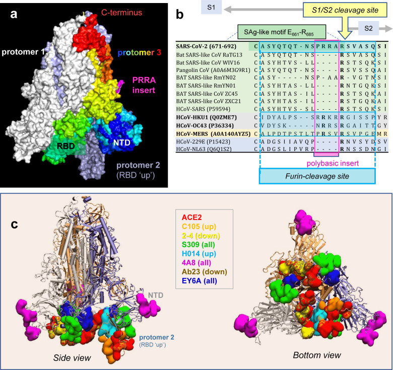
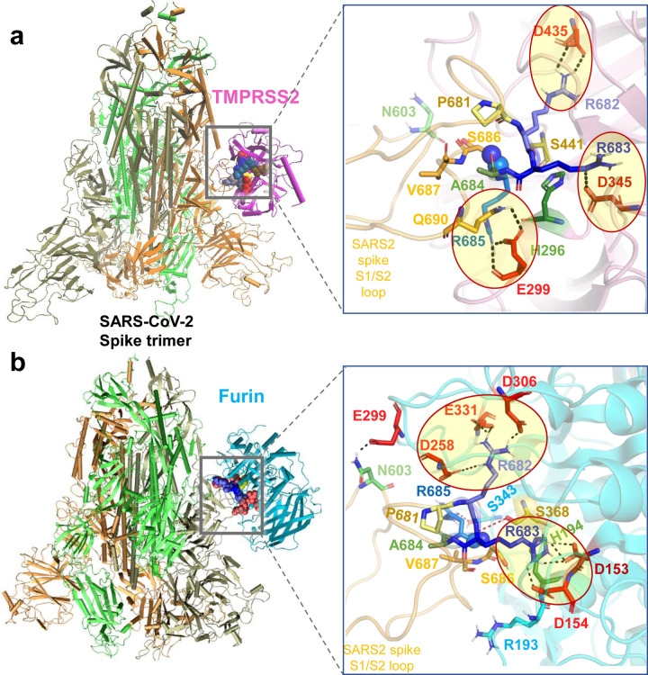
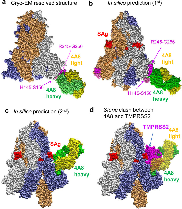
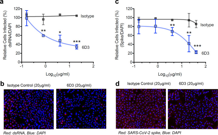
We recently discovered a superantigen-like motif, similar to Staphylococcal enterotoxin B (SEB), near the S1/S2 cleavage site of SARS-CoV-2 Spike protein, which might explain the multisystem-inflammatory syndrome (MIS-C) observed in children and cytokine storm in severe COVID-19 patients. We show here that an anti-SEB monoclonal antibody (mAb), 6D3, can bind this viral motif, and in particular its PRRA insert, to inhibit infection by blocking the access of host cell proteases, TMPRSS2 or furin, to the cleavage site. The high affinity of 6D3 for the furin-cleavage site originates from a poly-acidic segment at its heavy chain CDR2, a feature shared with SARS-CoV-2-neutralizing mAb 4A8. The affinity of 6D3 and 4A8 for this site points to their potential utility as therapeutics for treating COVID-19, MIS-C, or common cold caused by human coronaviruses (HCoVs) that possess a furin-like cleavage site.


Conflict of interest statement

Declarations of Interest

The authors have nothing to declare.

Figures

Figure 1:
Figure 1:. SARS-CoV-2 Spike (S) glycoprotein structure, sequence alignment against other CoVs, and interaction sites observed in cryo-EM studies with neutralizing antibodies.
(a) SARS-CoV-2 S trimer in the pre-fusion state. Protomers 1 and 2 are in white and light blue, respectively; and protomer 3, in spectral colors from blue (N-terminal domain, NTD; residue 1–305) to red (C-terminus), except for the 681PRRA684 insert in magenta. The insert is modeled using SWISS-MODEL. Each protomer’s RBD (residues 331–524) can assume up or down conformations as indicated, linked to the respective receptor-bound and -unbound states. (b) Sequence alignment of SARS-CoV-2 near the S1/S2 cleavage site against multiple bat and pangolin SARS-related strains, and other HCoVs, adjusted following previous studies,. Viruses belonging to the same lineage are shown by the same color shade; and HCoVs that encode furin-like cleavage sites are highlighted in bold fonts. Note that the polybasic insert PRRA of SARS-CoV-2 S is not found in closely related SARS-like CoVs but exists in MERS and HCoVs HKU1 and OC43. The furin-like cleavage site is indicated by the blue-shaded box. (c-d) Side (left) and bottom (right) views of receptor (ACE2)- and antibody-binding sites observed in cryo-EM structures resolved for the S protein complexed with the ACE2 and/or various Abs. The S trimer is shown in cartoons with the light blue protomer in the RBD-up conformation, and gray and light orange protomers in the RBD-down conformation. Binding sites for ACE2 and antibodies C10522, 2–423, S30924, H01425, 4A826, Ab2327, and EY6A are shown in space-filling surfaces in different colors (see the code in the inset). See Table 1 for additional information.
Figure 2:
Figure 2:. Binding poses of human proteases TMPRSS2 and furin to SARS-CoV-2 S protein.
(a-b) Structural models for the SARS-CoV-2 S protein complexed with TMPRSS2 (a), and furin (b), obtained from docking simulations followed by refinements. An overview (left) and a zoomed in view (right) are shown in each case. The arginines in the S1/S2 loop P681RRARS686 are shown in different shades of blue, and their interaction partner (acidic residues) in the proteases are shown in red. Spheres (right panels) highlight the peptide bond that would be cleaved (between R685 and S686). TMPRSS2 catalytic triad residues are in yellow for S441, green for H296, and dark red for D345. Their counterparts in furin are S368, H194 and D153. Note the short distance between the carbonyl carbon from R685 and the hydroxyl oxygen of the catalytic serine S441 of TMPRSS2 (3.5 Å) or S368 of furin (3.1 Å). Black dashed lines show the interfacial polar contacts and salt bridges, and those including the S1/S2 loop arginines are highlighted by ellipses.
Figure 3:
Figure 3:. Examination of binding characteristics of SARS-CoV-2-neutralizing mAbs 4A8.
(a) Cryo-EM structure (PDB: 7C2L); (b-c) Energetically most favorable conformers predicted for the S protein-4A8 complex. The former resembles the cryo-EM structure, involving the same segments, H145-S150 and R245-G256, at the binding epitopes of S. In the latter case the viral SAg-like region which also overlaps with the S1/S2 cleavage site serves as 4A8-binding epitope. (d) Competition between 4A8 and TMRPSS2 for binding to the S1/S2 cleavage site, based on the overlap between the binding poses of the two substrates. The diagram is generated by superposing the S-protein of the two complexes predicted in silico. The Spike-4A8 complex is generated in silico using the SARS-CoV-2 S structure with one RBD in the up conformer (PDB: 6VSB).
Figure 4:
Figure 4:. SEB-associated mAb 6D3 exhibits a high affinity to bind to the furin-cleavage site of SARS-CoV-2 S protein, potentially interfering with the S1/S2 cleavage by furin or TMPRSS2.
(a) Binding pose of three SEB-neutralizing Abs (mAbs 6D3, 14G8, and 20B1) onto SEB. The diagram is generated by superposing the crystal structures (PDB IDs 4RGN and 4RGM) resolved for the complexes. SEB is colored beige, with its SAg motif 150TNKKKATVQELD161 highlighted in blue space-filling. (b) Interface between 6D3 and SEB SAg motif. Heavy and light chains of 6D3 are colored green and cyan, respectively. Overall (c) and close-up (d) views of the complex formed between the S protein and anti-SEB mAb 6D3 and corresponding interfacial interactions engaging the arginines in the PRRA insert. SARS-CoV-2 S interfacial residues include I210-Q218, N603-Q607, E654-Y660 and A688-I693, and the SAg motif residues Y674, T678-R683. 6D3 interfacial residues include A24-K33, E50, D52, S54, D55, Y57, N59, K74-T77, and A100-A104 in the heavy chain, and D1, I2, Q27, N31-F38, Y55, W56, and D97-Y100 in the light chain. The Spike-4A8 complex is generated in silico using SARS-CoV-2 S structure with one RBD in the up conformer (PDB: 6VSB).
Figure 5:
Figure 5:. Monoclonal antibody 6D3 prevents SARS-CoV-2 infection.
6D3 or isotype control antibodies (at indicated concentrations) were incubated with virus (100 PFU/well) for 1 hour at room temperature before addition to Vero-E6 cells (5×10³ cells/well). 48 hours post infection cells were fixed and stained for dsRNA or SARS-CoV-2 spike protein. (a) Quantification of the percentage of infected cells per well by dsRNA staining. Data are representative of two independent experiments. (b) Representative fluorescence images of 6D3-mediated inhibition of virus infection (dsRNA). (c) Quantification of the percentage of infected cells per well by spike staining. (d) Representative fluorescence images of 6D3-mediated inhibition of virus infection (spike). Data were analyzed by t test (6D3 vs. isotype control) with multiple testing correction (FDR). See also Extended Data Fig. 3 for detailed results as a function of 6D3 concentration.
Figure 6:
Figure 6:. Polyacidic residues in the CDR2 of the mAbs 6D3 and 4A8 heavy chains play a major role in blocking the furin-like cleavage site of HCoV S protein.
(a) Multiple sequence alignment of the VH domain of anti-SEB Abs (6D3, 14G8 and 20B1) and anti-SARS-CoV-2 S Abs (listed in the left column). Using Ab 4A8 as the reference, the residue ranges of the three CDRs are: CDR1 (residues 25 to 32), CDR2 (51 to 58), and CDR3 (100 to 116), as indicated by the blue bars. (b) Overall and close-up views of the complex and interfacial interaction of SARS-CoV-2 S protein and 6D3 antibody. Note that three acidic residues from CDR2 interact with the basic residues R682, R683 and R685 of the S protein. (c) Same as b, repeated for 4A8. A cluster of acidic residues from CDR2 forms a network of interactions with the S protein R682, R683 and R685. The complexes in panels b and c are generated in silico using SARS-CoV-2 S structure with three RBDs in the down conformer (PDB: 6VXX). The residues belonging to the Abs are labelled in lightface, those of the S protein in boldface. HCoV-OC43 encodes a S1/S2 furin-like cleavage site at 754RRARG758. (d) Overall and close-up views of the complex formed, and interfacial interactions, between the HCoV-OC43 S protein (with S1/S2 furin-like cleavage site 754RRARG758) and the Ab 6D3 (top) and 4A8 (bottom). Note that three acidic residues from CDR2 interact with R754, R755 and R757 in hCoV-OC43 S protein. The residues belonging to the Abs are labelled in lightface, those of the HCoV in boldface.

References

    1. Tay M.Z., Poh C.M., Rénia L., MacAry P.A. & Ng L.F.P. The trinity of COVID-19: immunity, inflammation and intervention. Nat. Rev. Immunol. 20, 363–374 (2020). - PMC - PubMed
    1. Vabret N. et al. Immunology of COVID-19: current state of the science. Immunity 52, 910–941 (2020). - PMC - PubMed
    1. Gupta A. et al. Extrapulmonary manifestations of COVID-19. Nat Med 26, 1017–1032 (2020). - PMC - PubMed
    1. Riphagen S., Gomez X., Gonzalez-Martinez C., Wilkinson N. & Theocharis P. Hyperinflammatory shock in children during COVID-19 pandemic. Lancet 395, 1607–1608 (2020). - PMC - PubMed
    1. Verdoni L. et al. An outbreak of severe Kawasaki-like disease at the Italian epicentre of the SARS-CoV-2 epidemic: an observational cohort study. Lancet 395, 1771–1778 (2020). - PMC - PubMed

Publication types