

Tongue and hypoglossal morphology after intralingual cholera toxin B-saporin injection
- PMID: 33269488
- PMCID: PMC8117177
- DOI: 10.1002/mus.27131
Tongue and hypoglossal morphology after intralingual cholera toxin B-saporin injection
Abstract
Introduction: We recently developed an inducible model of dysphagia using intralingual injection of cholera toxin B conjugated to saporin (CTB-SAP) to cause death of hypoglossal neurons. In this study we aimed to evaluate tongue morphology and ultrastructural changes in hypoglossal neurons and nerve fibers in this model.
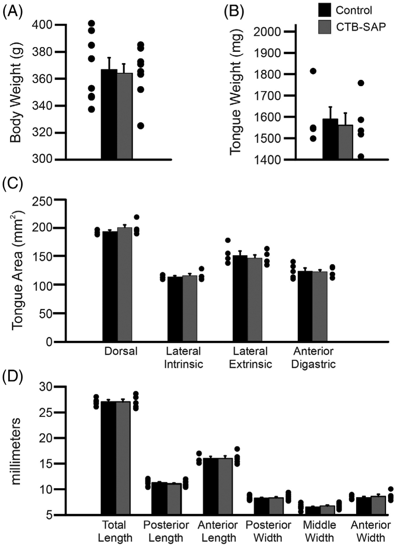
Methods: Tissues were collected from 20 rats (10 control and 10 CTB-SAP animals) on day 9 post-injection. Tongues were weighed, measured, and analyzed for microscopic changes using laminin immunohistochemistry. Hypoglossal neurons and axons were examined using transmission electron microscopy.
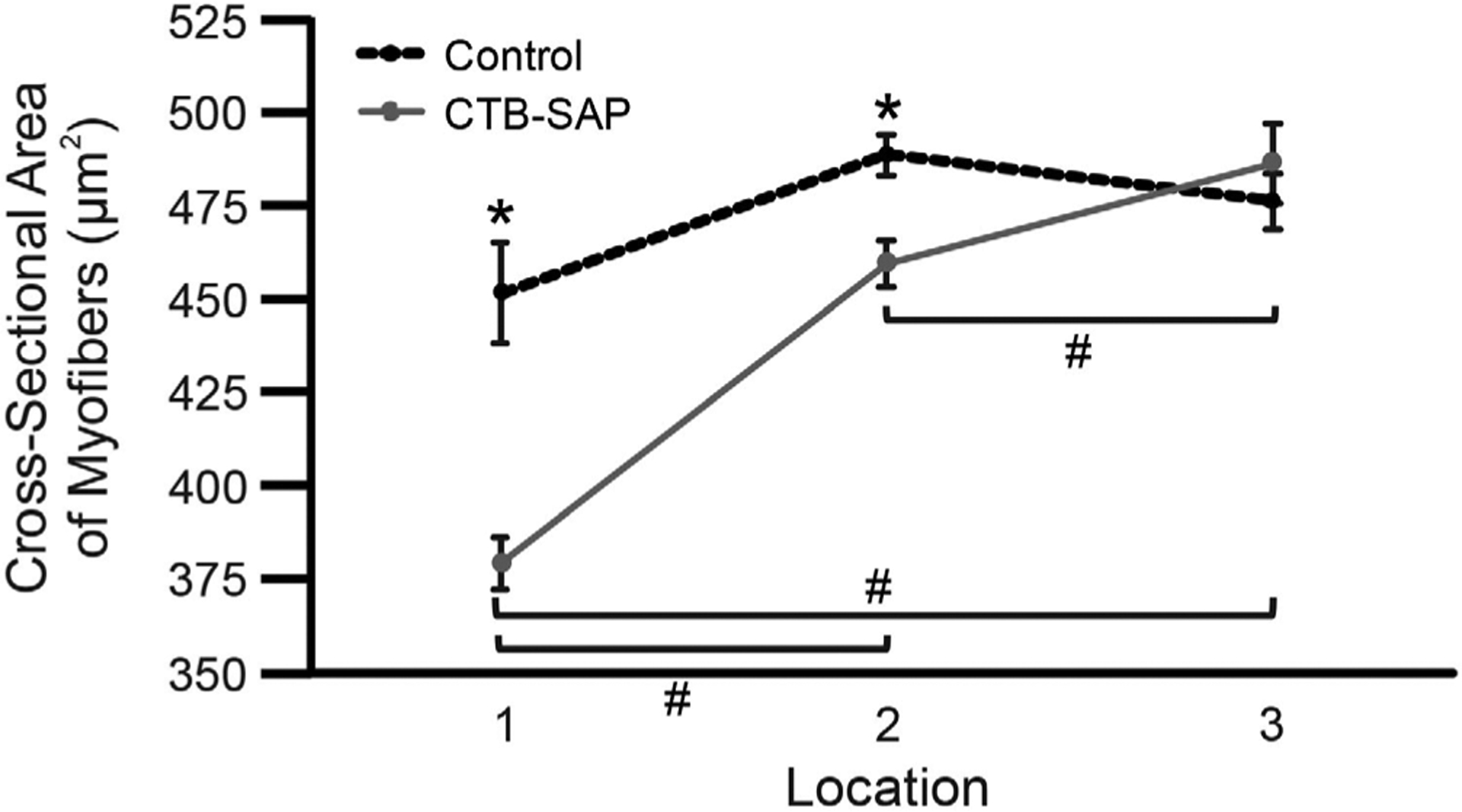
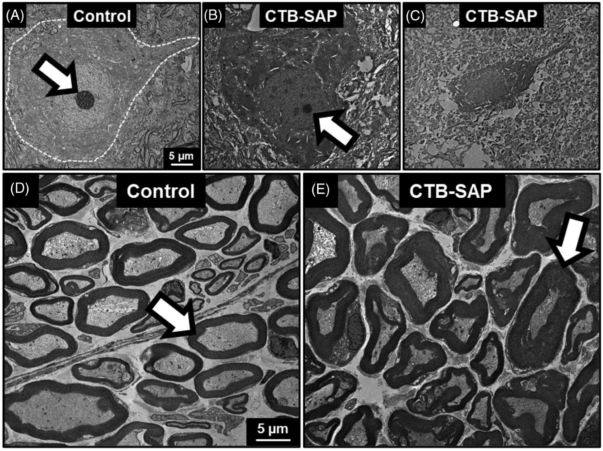
Results: The cross-sectional area of myofibers in the posterior genioglossus was decreased in CTB-SAP-injected rats. Degenerative changes were observed in both the cell bodies and distal axons of hypoglossal neurons.
Discussion: Preliminary results indicate this model may have translational application to a variety of neurodegenerative diseases resulting in tongue dysfunction and associated dysphagia.
Keywords: denervation atrophy, dysphagia, motor neuron death, rodent models, ultrastructural changes.
© 2020 Wiley Periodicals LLC.
Conflict of interest statement
CONFLICTS OF INTEREST
The authors declare no potential conflicts of interest.
Figures
Similar articles
-
A Strength Endurance Exercise Paradigm Mitigates Deficits in Hypoglossal-Tongue Axis Function, Strength, and Structure in a Rodent Model of Hypoglossal Motor Neuron Degeneration.Front Neurosci. 2022 Jun 30;16:869592. doi: 10.3389/fnins.2022.869592. eCollection 2022. Front Neurosci. 2022. PMID: 35844238 Free PMC article.
-
Hypoglossal Motor Neuron Death Via Intralingual CTB-saporin (CTB-SAP) Injections Mimic Aspects of Amyotrophic Lateral Sclerosis (ALS) Related to Dysphagia.Neuroscience. 2018 Oct 15;390:303-316. doi: 10.1016/j.neuroscience.2018.08.026. Epub 2018 Sep 1. Neuroscience. 2018. PMID: 30179644 Free PMC article.
-
Respiratory function after selective respiratory motor neuron death from intrapleural CTB-saporin injections.Exp Neurol. 2015 May;267:18-29. doi: 10.1016/j.expneurol.2014.11.011. Epub 2014 Dec 2. Exp Neurol. 2015. PMID: 25476493 Free PMC article.
-
Two types of inhibitory postsynaptic potentials in the hypoglossal motoneurons.Prog Neurobiol. 1993 Mar;40(3):385-411. doi: 10.1016/0301-0082(93)90016-l. Prog Neurobiol. 1993. PMID: 8441813 Review. No abstract available.
-
Gene delivery to the hypoglossal motor system: preclinical studies and translational potential.Gene Ther. 2021 Aug;28(7-8):402-412. doi: 10.1038/s41434-021-00225-1. Epub 2021 Feb 11. Gene Ther. 2021. PMID: 33574581 Free PMC article. Review.
Cited by
-
Impact of Limb Phenotype on Tongue Denervation Atrophy, Dysphagia Penetrance, and Survival Time in a Mouse Model of ALS.Dysphagia. 2022 Dec;37(6):1777-1795. doi: 10.1007/s00455-022-10442-4. Epub 2022 Apr 15. Dysphagia. 2022. PMID: 35426522 Free PMC article.
-
A Strength Endurance Exercise Paradigm Mitigates Deficits in Hypoglossal-Tongue Axis Function, Strength, and Structure in a Rodent Model of Hypoglossal Motor Neuron Degeneration.Front Neurosci. 2022 Jun 30;16:869592. doi: 10.3389/fnins.2022.869592. eCollection 2022. Front Neurosci. 2022. PMID: 35844238 Free PMC article.
-
Tongue exercise ameliorates structural and functional upper airway deficits in a rodent model of hypoglossal motor neuron loss.Front Neurol. 2024 Sep 4;15:1441529. doi: 10.3389/fneur.2024.1441529. eCollection 2024. Front Neurol. 2024. PMID: 39296960 Free PMC article.
-
Saporin as a Commercial Reagent: Its Uses and Unexpected Impacts in the Biological Sciences-Tools from the Plant Kingdom.Toxins (Basel). 2022 Mar 2;14(3):184. doi: 10.3390/toxins14030184. Toxins (Basel). 2022. PMID: 35324681 Free PMC article. Review.
-
Axon and Myelin Sheath Segmentation in Electron Microscopy Images using Meta Learning.IEEE Appl Imag Pattern Recognit Workshop. 2022 Oct;2022:10.1109/aipr57179.2022.10092238. doi: 10.1109/aipr57179.2022.10092238. Epub 2023 Apr 10. IEEE Appl Imag Pattern Recognit Workshop. 2022. PMID: 37214276 Free PMC article.
References
-
- Bruijn LI, Miller TM, Cleveland DW. Unraveling the mechanisms involved in motor neuron degeneration in ALS. Annu Rev Neurosci. 2004;27:723–749. - PubMed
-
- Gonzalez de Aguilar JL, Echaniz-Laguna A, Fergani A, et al. Amyotrophic lateral sclerosis: all roads lead to Rome. J Neurochem. 2007;101:1153–1160. - PubMed
-
- Brooks BR, Miller RG, Swash M, Munsat TL, World Federation of Neurology Research Group on Motor Neuron Diseases. El Escorial revisited: revised criteria for the diagnosis of amyotrophic lateral sclerosis. Amyotroph Lateral Scler Other Motor Neuron Disord. 2000;1: 293–299. - PubMed
Publication types
MeSH terms
Substances
Grants and funding
LinkOut - more resources
Full Text Sources
Medical
Miscellaneous
