. 2021 May-Jun;23(3):306-313.
doi: 10.4103/aja.aja_76_20.

A decrease of docosahexaenoic acid in testes of mice fed a high-fat diet is associated with impaired sperm acrosome reaction and fertility

Affiliations

A decrease of docosahexaenoic acid in testes of mice fed a high-fat diet is associated with impaired sperm acrosome reaction and fertility

Julio Bunay et al. Asian J Androl. 2021 May-Jun.

Abstract

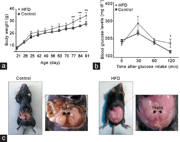
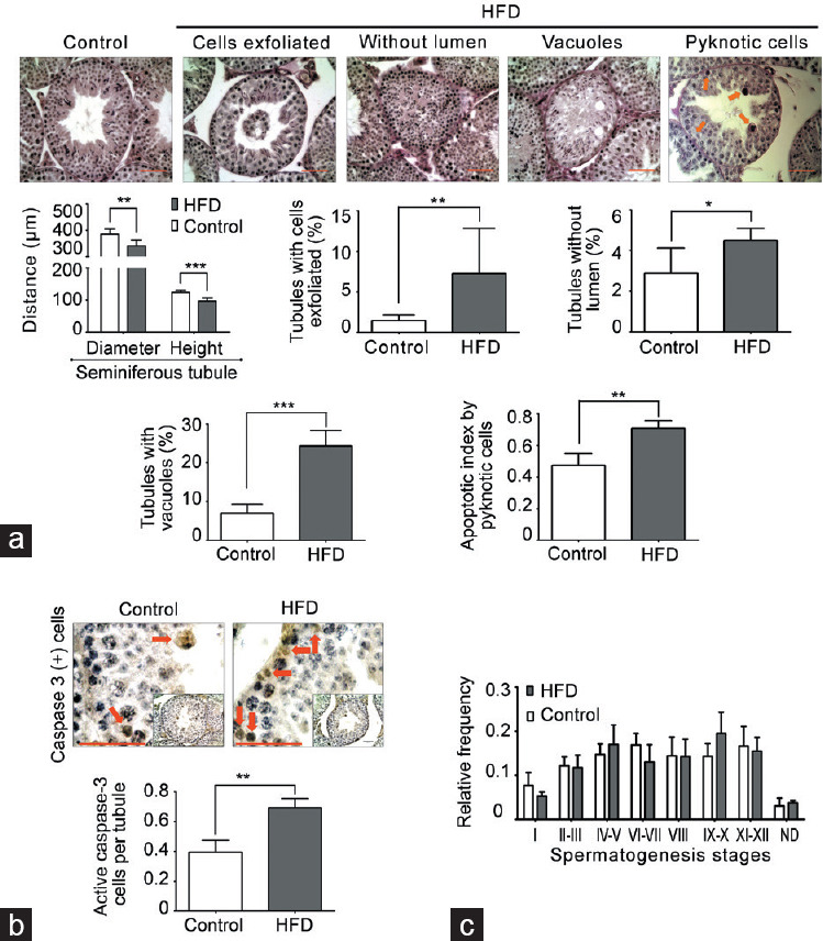
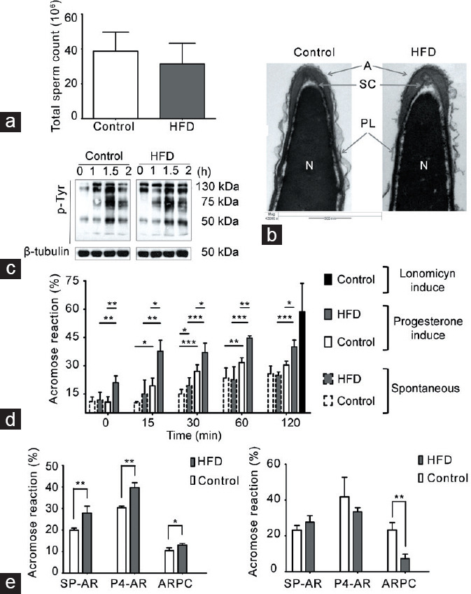
Obesity is a major worldwide health problem that is related to most chronic diseases, including male infertility. Owing to its wide impact on health, mechanisms underlying obesity-related infertility remain unknown. In this study, we report that mice fed a high-fat diet (HFD) for over 2 months showed reduced fertility rates and increased germ cell apoptosis, seminiferous tubule degeneration, and decreased intratesticular estradiol (E2) and E2-to-testosterone ratio. Interestingly, we also detected a decrease in testicular fatty acid levels, behenic acid (C22:0), and docosahexaenoic acid (DHA, 22:6n-3), which may be related to the production of dysfunctional spermatozoa. Overall, we did not detect any changes in the frequency of seminiferous tubule stages, sperm count, or rate of in vitro capacitation. However, there was an increase in spontaneous and progesterone-induced acrosomal exocytosis (acrosome reaction) in spermatozoa from HFD-fed mice. These data suggest that a decrease in E2 and fatty acid levels influences spermatogenesis and some steps of acrosome biogenesis that will have consequences for fertilization. Thus, our results add new evidence about the adverse effect of obesity in male reproduction and suggest that the acrosomal reaction can also be affected under this condition.

Keywords: cholesterol; estradiol; fat acid; testis; testosterone.


Conflict of interest statement

None

Figures

Figure 1
Figure 1
HFD feeding induced a metabolic state associated with alterations in reproductive organs. Metabolic evaluation of HFD-fed compared with chow-diet (control) mice: (a) body weight and (b) glucose tolerance test. (c) Representative images of accumulation of fatty tissue in the abdominal cavity of HFD-fed mice. All graphs represent the mean ± s.d., n = 4. Data were statistically analyzed with an unpaired t-test: *P < 0.05, **P < 0.01. s.d.: standard deviation; HFD: high-fat diet.
Figure 2
Figure 2
Degeneration-atrophy of seminiferous tubules and increase in apoptosis in HFD-fed mice. (a) Morphometric analysis of seminiferous tubules (diameter and epithelial height). In addition, images of testicular sections stained with PAS and hematoxylin that present different types of seminiferous tubule degeneration/atrophy and germ cell death by pyknotic cells (orange arrow) found in HFD-fed compared with chow-fed mice. The quantification of histological alterations and germ cell death in HFD-fed versus chow-fed mice is shown at the bottom. Scale bars = 100 μm. (b) Evaluation of germ cell apoptosis by caspase-3-positive cells (red arrow). Scale bars = 100 μm. (c) Frequency of seminiferous epithelial cycles. All graphs represent the mean ± s.d., n = 4. Data were statistically analyzed with an unpaired t-test: *P < 0.05, **P < 0.01, ***P < 0.001. s.d.: standard deviation; HFD: high-fat diet; PAS: periodic acid–Schiff.
Figure 3
Figure 3
A decrease in intratesticular estradiol (E2) levels was not related to changes in E2 synthesis in mice fed with a HFD. (a) Testosterone levels in serum and seminiferous tubular fluid (intratesticular). (b) E2 levels in serum and seminiferous tubular fluid (intratesticular). (c) E2-to-testosterone ratio in serum and seminiferous tubular fluid (intratesticular). All measurements were made by radioimmunoassay. All graphs represent the mean ± s.d., n = 4. Data were statistically analyzed with an unpaired t-test: *P < 0.05. (d) Cyp19a1 (aromatase) gene expression by quantitative polymerase chain reaction in adult mouse testes at the experimental end point. All graphs represent the mean ± s.d., n = 3. Data were analyzed by Wilcoxon signed-rank test. AU: arbitrary units; s.d.: standard deviation; HFD: high-fat diet.
Figure 4
Figure 4
Capacitation and the AR in spermatozoa from HFD-fed mice. Comparison between HFD-fed and chow-diet (control) mice. (a) Total sperm count in cauda epididymis, graph represents the mean ± s.d., n = 4. No significant differences were observed. (b) Representative images of acrosomal ultrastructure in HFD-fed and chow-fed mice. The images depict the acrosome (A), sub-acrosomal compartment (SC), nucleus (N), and plasma membrane (PL). (c) Representative images of the time-dependent pattern of tyrosine phosphorylation; β-tubulin was used as a loading control, n = 3. (d) Quantification of AR; spermatozoa from each group were incubated in CM. The time-dependent percentage of spontaneous AR (SP-AR) and AR induced by 40 μmol l−1 of progesterone (P4-AR) was evaluated with the Coomassie blue dye technique. As a control, the maximum AR response was evaluated in the presence of 10 μmol l−1 A23187, a calcium ionophore. All graphs represent the mean ± s.d., n = 4. SP-AR and P4-AR were analyzed with an unpaired t-test: *P < 0.05, **P < 0.01, ***P < 0.001. (e) SP-AR and P4-AR and the rate of ARPC were measured with the Coomassie blue dye technique, after incubating the spermatozoa in CM with 10 mg ml−1 or 30 mg ml−1 bovine serum albumin as a cholesterol scavenger. All graphs represent the mean ± s.d., n = 4. Data were statistically analyzed with an unpaired t-test: *P < 0.05, **P < 0.01. s.d.: standard deviation; AR: acrosomal reaction; HFD: high-fat diet; CM: capacitating medium; ARPC: AR after progesterone challenger.

References

    1. Esteves SC, Hamada A, Kondray V. What every gynecologist should know about male infertility: an update. Arch Gynecol Obstet. 2012;286:217–29. - PubMed
    1. NCD Risk Factor Collaboration. Worldwide trends in body-mass index, underweight, overweight, and obesity from 1975 to 2016: a pooled analysis of 2416 population-based measurement studies in 128.9 million children, adolescents, and adults. Lancet. 2017;390:2627–42. - PMC - PubMed
    1. Pedro J, Brandão T, Schmidt L, Costa ME, Martins MV. What do people know about fertility.A systematic review on fertility awareness and its associated factors? Ups J Med Sci. 2018;123:71–81. - PMC - PubMed
    1. Reis LO, Dias FG. Male fertility, obesity, and bariatric surgery. Reprod Sci. 2012;8:778–85. - PubMed
    1. Abiad F, Awwad J, Abbas HA, Zebian D, Ghazeeri G. Management of weight loss in obesity-associated male infertility: a spotlight on bariatric surgery. Human Fertil (Camb) 2017;20:227–35. - PubMed

Publication types

Substances