. 2021 Mar 5;16(3):302-314.
doi: 10.1093/scan/nsaa165.

Stereotypes bias face perception via orbitofrontal-fusiform cortical interaction

Affiliations

Stereotypes bias face perception via orbitofrontal-fusiform cortical interaction

Benjamin O Barnett et al. Soc Cogn Affect Neurosci. .

Abstract

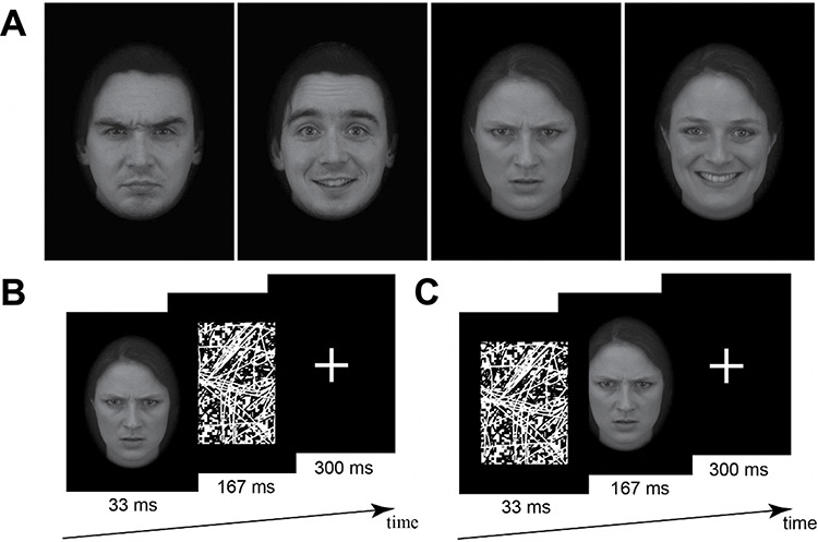
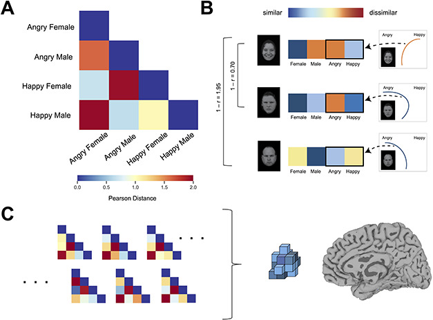
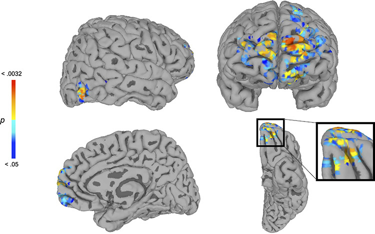
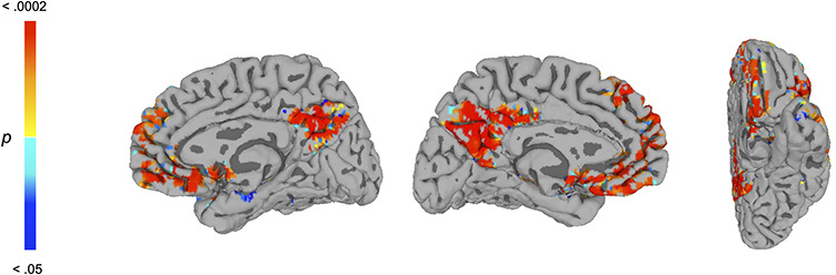
Previous research has shown that social-conceptual associations, such as stereotypes, can influence the visual representation of faces and neural pattern responses in ventral temporal cortex (VTC) regions, such as the fusiform gyrus (FG). Current models suggest that this social-conceptual impact requires medial orbitofrontal cortex (mOFC) feedback signals during perception. Backward masking can disrupt such signals, as it is a technique known to reduce functional connectivity between VTC regions and regions outside VTC. During functional magnetic resonance imaging (fMRI), subjects passively viewed masked and unmasked faces, and following the scan, perceptual biases and stereotypical associations were assessed. Multi-voxel representations of faces across the VTC, and in the FG and mOFC, reflected stereotypically biased perceptions when faces were unmasked, but this effect was abolished when faces were masked. However, the VTC still retained the ability to process masked faces and was sensitive to their categorical distinctions. Functional connectivity analyses confirmed that masking disrupted mOFC-FG connectivity, which predicted a reduced impact of stereotypical associations in the FG. Taken together, our findings suggest that the biasing of face representations in line with stereotypical associations does not arise from intrinsic processing within the VTC and FG alone, but instead it depends in part on top-down feedback from the mOFC during perception.

Keywords: face perception; multivariate fMRI; social cognition; social vision; stereotypes.


Figures

Fig. 1.
Fig. 1.
(A) Example face stimuli. Examples of angry male, happy male, angry female and happy female face stimuli used in the fMRI, discrimination and mouse-tracking tasks. fMRI procedure. (B) Masked condition and (C) unmasked condition. In both, each sequence was repeated four times within a trial (totaling 2000 ms).
Fig. 2.
Fig. 2.
(A) The group-average subjective DM. Warmer colors represent greater dissimilarity. (B) The method by which a subject’s subjective DM was computed. In this example (Angry Male condition), MD values for the four categories (Female, Male, Angry and Happy), which reflect mouse trajectories’ attraction toward the four category responses in the mouse-tracking tasks, were used to create a response vector. Similarity in these response vectors was used to calculate pairwise similarity for the other conditions (Happy Female, Angry Female and Angry Male). In this way, face conditions that elicited similar activation of the Female, Male, Angry and Happy responses during mouse-tracking were deemed more similar in a subject’s subjective DM. (C) An illustration of our RSA procedure. Participants’ neural patterns for each condition were correlated with each other to formulate a neural DM. A subject’s own subjective DM was used to predict this neural DM (when controlling for the group-average subjective DM depicted in A).
Fig. 3.
Fig. 3.
Whole-brain searchlight RSA results in the unmasked condition, revealing the right FG and mOFC. At each searchlight sphere, subjects’ own subjective DM was used to predict neural-pattern structure, while controlling for the group-average subjective DM. This analysis revealed regions in the right FG (top left), mOFC (bottom) and mPFC (top right), showing that these regions’ neural-pattern structure reflected subjects’ stereotypically biased subjective perceptions, even when any common bias shared across subjects or intrinsic to the stimuli are accounted for (P < 0.05, corrected). No regions survived correction for this analysis in the masked condition (P < 0.05, corrected).
Fig. 4.
Fig. 4.
Whole-brain PPI analysis eliciting regions whose functional connectivity with the rFG seed region was modulated by masked vs unmasked conditions. A PPI analysis was conducted using a face-sensitive region of the right FG as a seed, revealing an extensive portion of the mOFC and additional regions involved in social cognition, including the mPFC (P < 0.05, corrected). These regions showed diminished functional connectivity with the right FG when subjects viewed masked as compared with unmasked faces. See Table 1 for list of regions.

References

    1. Adams R.B., Ambady N., Nakayama K., Shimojo S. (2011). The Science of Social Vision. New York: Oxford University Press.
    1. Amodio D.M. (2014). The neuroscience of prejudice and stereotyping. Nature Reviews: Neuroscience, 15(10), 670–82. - PubMed
    1. Avants B.B., Epstein C.L., Grossman M., Gee J.C. (2008). Symmetric diffeomorphic image registration with cross-correlation: evaluating automated labeling of elderly and neurodegenerative brain. Medical Image Analysis, 12(1), 26–41. - PMC - PubMed
    1. Baars B.J., Franklin S., Ramsoy T.Z. (2013). Global workspace dynamics: cortical “binding and propagation” enables conscious contents. Frontiers in Psychology, 4, 200. - PMC - PubMed
    1. Bagnis A., Celeghin A., Diano M., et al. (2020). Functional neuroanatomy of racial categorization from visual perception: a meta-analytic study. Neuroimage, 207, 116939. - PubMed

Publication types