Observational Study
. 2020 Dec 3;15(12):e0243346.
doi: 10.1371/journal.pone.0243346. eCollection 2020.

Retinal changes in COVID-19 hospitalized cases

Affiliations
Observational Study

Retinal changes in COVID-19 hospitalized cases

Rafael Lani-Louzada et al. PLoS One. .

Abstract

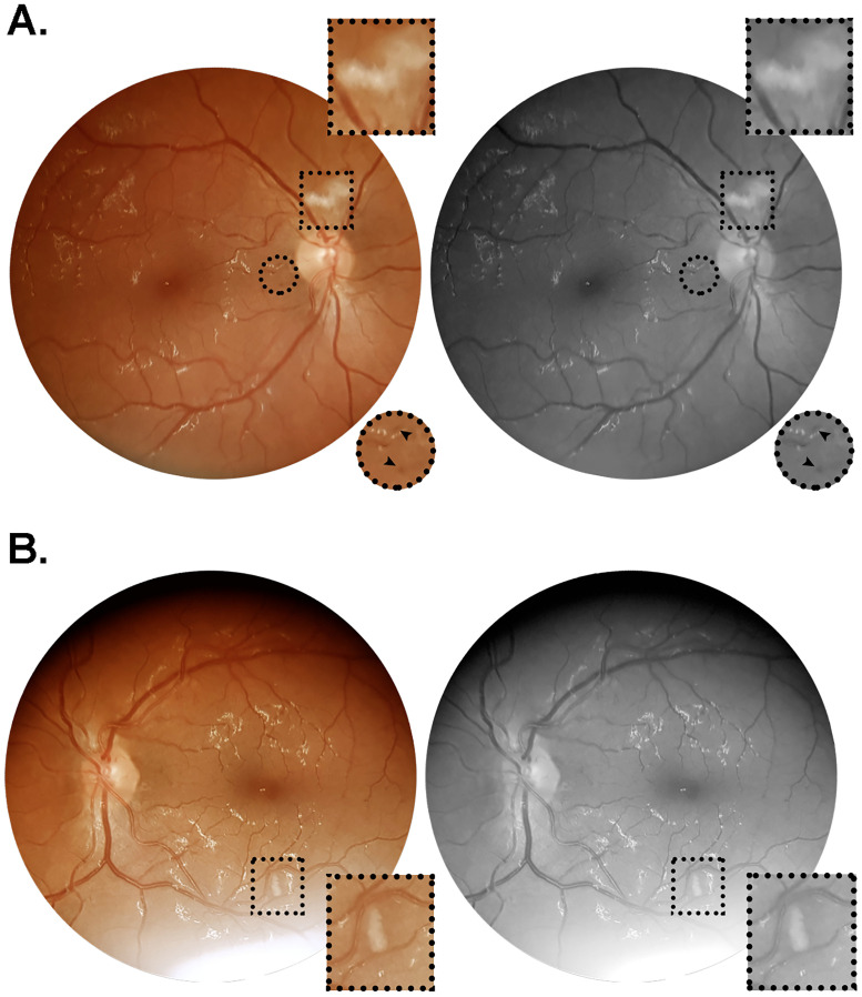
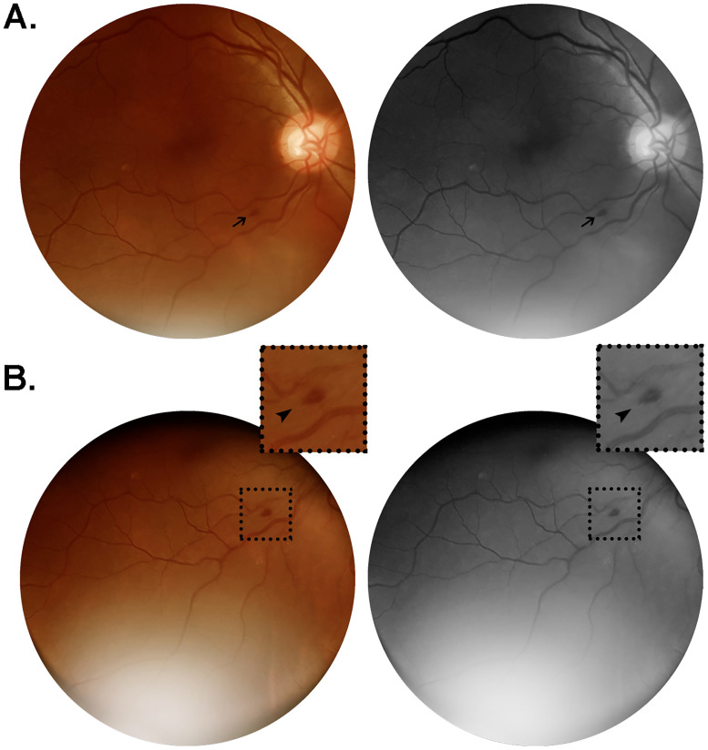
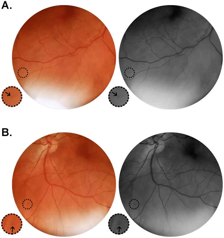
The main objective of this study was to evaluate the retinas of severely or critically ill COVID-19 patients during their hospital stay, at varying time points after symptoms onset. This was a case series observed during May 2020 in two referral centers for COVID-19 treatment in Rio de Janeiro, Brazil. 47 eyes from 25 hospitalized patients with severe or critical confirmed illness were evaluated. A handheld retinal camera was used to acquire bilateral fundus images at several time points after symptoms onset. Electronic health records were retrospectively analyzed and clinical data collected. Severe and critical diseases were noticed in 52% (13/25) and 48% (12/25) of enrolled patients, respectively. Retinal changes were present in 12% (3/25) of patients: a 35 year-old male demonstrated bilateral nerve fiber layer infarcts and microhemorrhages in the papillomacular bundle, but required mechanical ventilation and developed severe anemia and systemic hypotension, acute kidney injury and neurologic symptoms during the course of the disease (critical illness); a 56 year-old male, who required full enoxaparin anticoagulation due to particularly elevated D-dimer (>5.0 mcg/mL), demonstrated unilateral and isolated flame-shaped hemorrhages; and a 49 year-old hypertensive male showed bilateral and discrete retinal dot and blot microhemorrhages. The other 22 patients evaluated did not demonstrate convincing retinal changes upon examination. There was no correlation between disease severity and admission serum levels of CRP, D-dimer and ferritin. This was the first study to show that vascular retinal changes may be present in not insignificant numbers of severe or critical COVID-19 inpatients. These retinal changes, only seen after morbid developments, were likely secondary to clinical intercurrences or comorbidities instead of a direct damage by SARS-CoV-2, and may be important and easily accessible outcome measures of therapeutic interventions and sentinels of neurologic and systemic diseases during COVID-19 pandemic.


Conflict of interest statement

The authors have declared that no competing interests exist.

Figures

Fig 1
Fig 1. Color and red-free retinal photographs from patient #6 (W.A.C.).
Images acquired on day 64 after symptoms onset, from a 35 year-old obese and hypertensive man who was admitted on day seven since onset of fever, cough and dyspnea. During hospitalization, this patient evolved to critical illness, characterized by acute respiratory distress syndrome, respiratory failure and acute kidney injury, requiring ICU monitorization, mechanical ventilation and hemodialysis. The patient had a positive clinical outcome, and received hospital discharge on day 69 after symptoms onset. (A) Right retina showing a nerve fiber layer infarct above the optic nerve head, and microhemorrhages in the papillomacular bundle close to the optic disc. (B) Left retina showing nerve fiber layer infarcts at the inferior temporal vascular arcade, approximately 1.5 disc diameters inferior to the macula. Square and circular insets represent 2x magnification of the delimited area.
Fig 2
Fig 2. Color and red-free retinal photographs from patient #18 (W.S.).
Images acquired on day 16 from symptoms onset, from a 56 year-old man with no comorbidities, admitted on day 13 after onset of a clinical scenario characterized by fever, cough, headache, myalgia and dyspnea. The patient evolved to severe illness, not requiring ICU monitorization nor developing any organ failure. Lung CT at admission revealed bilateral ground-glass opacities predominantly in the periphery, extending to approximately 50% of lung parenchyma. (A) Flame-shaped hemorrhage (arrow) approximately one disc diameter from the optic nerve head, close to the inferior temporal vascular arcade. (B) Same lesion observed from an alternative point of gaze fixation, magnified in the insets (2x; arrowheads).
Fig 3
Fig 3. Color and red-free retinal photographs from patient #19 (G.C.X.).
Images acquired on day 12 from symptoms onset, from a 49 year-old man in regular treatment for arterial hypertension, admitted on the eight day after onset of fever, cough, myalgia and progressive dyspnea. Lung CT on admission showed typical viral pneumonia findings extending through 25–50% of the parenchyma. The patient evolved to a severe phenotype of the disease. (A) Right retina presenting with isolated microhemorrhage at the periphery close to the inferior temporal vasculature. (B) Left retina showing isolated microhemorrhage at the inferior nasal retinal quadrant. Insets represent delimited areas with 2x magnification.

References

    1. Coronavirus disease (COVID-19) [Internet]. [cited 2020 Aug 29]. https://www.who.int/emergencies/diseases/novel-coronavirus-2019?gclid=Cj....
    1. Jiang F, Deng L, Zhang L, Cai Y, Cheung CW, Xia Z. Review of the Clinical Characteristics of Coronavirus Disease 2019 (COVID-19). J Gen Intern Med. 2020;35(5):1545–9. 10.1007/s11606-020-05762-w - DOI - PMC - PubMed
    1. Huang C, Wang Y, Li X, Ren L, Zhao J, Hu Y, et al. Clinical features of patients infected with 2019 novel coronavirus in Wuhan, China. Lancet. 2020;395(10223):497–506. 10.1016/S0140-6736(20)30183-5 - DOI - PMC - PubMed
    1. Fotuhi M, Mian A, Meysami S, Raji CA. Neurobiology of COVID-19. J Alzheimer’s Dis. 2020;76:1–17. 10.3233/JAD-200581 - DOI - PMC - PubMed
    1. Zubair AS, McAlpine LS, Gardin T, Farhadian S, Kuruvilla DE, Spudich S. Neuropathogenesis and Neurologic Manifestations of the Coronaviruses in the Age of Coronavirus Disease 2019: A Review. JAMA Neurol. 2020;1–10. 10.1001/jamaneurol.2020.2065 - DOI - PMC - PubMed

Publication types