. 2020 Dec 3;16(12):e1009125.
doi: 10.1371/journal.ppat.1009125. eCollection 2020 Dec.

The cucumber mosaic virus 1a protein regulates interactions between the 2b protein and ARGONAUTE 1 while maintaining the silencing suppressor activity of the 2b protein

Affiliations

The cucumber mosaic virus 1a protein regulates interactions between the 2b protein and ARGONAUTE 1 while maintaining the silencing suppressor activity of the 2b protein

Lewis G Watt et al. PLoS Pathog. .

Abstract

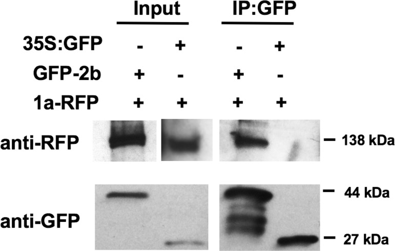
The cucumber mosaic virus (CMV) 2b viral suppressor of RNA silencing (VSR) is a potent counter-defense and pathogenicity factor that inhibits antiviral silencing by titration of short double-stranded RNAs. It also disrupts microRNA-mediated regulation of host gene expression by binding ARGONAUTE 1 (AGO1). But in Arabidopsis thaliana complete inhibition of AGO1 is counterproductive to CMV since this triggers another layer of antiviral silencing mediated by AGO2, de-represses strong resistance against aphids (the insect vectors of CMV), and exacerbates symptoms. Using confocal laser scanning microscopy, bimolecular fluorescence complementation, and co-immunoprecipitation assays we found that the CMV 1a protein, a component of the viral replicase complex, regulates the 2b-AGO1 interaction. By binding 2b protein molecules and sequestering them in P-bodies, the 1a protein limits the proportion of 2b protein molecules available to bind AGO1, which ameliorates 2b-induced disease symptoms, and moderates induction of resistance to CMV and to its aphid vector. However, the 1a protein-2b protein interaction does not inhibit the ability of the 2b protein to inhibit silencing of reporter gene expression in agroinfiltration assays. The interaction between the CMV 1a and 2b proteins represents a novel regulatory system in which specific functions of a VSR are selectively modulated by another viral protein. The finding also provides a mechanism that explains how CMV, and possibly other viruses, modulates symptom induction and manipulates host-vector interactions.


Conflict of interest statement

The authors have declared that no competing interests exist.

Figures

Fig 1
Fig 1. The effects on aphid reproduction of cucumber mosaic virus (CMV) infection and transgenic expression the CMV 1a and 2b proteins in Arabidopsis thaliana.
Individual one-day-old Myzus persicae nymphs (n≥16) were placed on plants and number of offspring (colony size) counted at 10 days post-infestation. Aphids were placed on plants that were: non-transgenic or transgenic plants constitutively expressing the CMV 2b protein (lines 2.30F and 3.13F [32]), the CMV 1a protein or both the 1a and 2b proteins [41]. Different letters are assigned to significantly different groups (ANOVA with post-hoc Tukey’s tests, P<0.05). Error bars represent standard error of the mean.
Fig 2
Fig 2. Sub-cellular localization of the cucumber mosaic virus 1a and 2b proteins.
The GFP-1a and RFP-1a protein fusions were expressed in N. benthamiana leaves by agroinfiltration and images recorded 3–4 days later by confocal laser scanning microscopy. Consistent with previous investigations of the Fny-CMV 2b protein [22,23], GFP-2b (A) and 2b-RFP (B) accumulated in the nuclei and cytoplasm. In contrast, GFP-1a (C) and RFP-1a (D) accumulated as punctate specks of varying size. At higher magnification (C, right panel) GFP-1a accumulation at the cell periphery could be observed. However, staining with a membrane-binding dye (FM-4-64) indicated that the larger GFP-1a aggregations did not co-localize with the cell membrane (E). Staining with ER-tracker (ER) did not indicate co-localization of GFP-1a with the endoplasmic reticulum network (F).
Fig 3
Fig 3. The P-body marker DCP1 localizes with the cucumber mosaic virus 1a protein.
A. Fluorescence from DCP1-GFP was observed as punctate specks with varying size. The localization pattern of DCP1-GFP resembled that of RFP-1a, leading us to speculate that they may occupy the same subcellular compartment. RFP-DCP1 and DCP1-GFP were observed in small foci associated with the periphery of the cell, presumably P-bodies. DCP1-GFP fluorescence was brighter than DCP1-RFP so was used for co-localization experiments with RFP-1a (Panel B). B. When RFP-1a was co-agroinfiltrated with DCP1-GFP we observed co-localization of the two proteins in some of the specks.
Fig 4
Fig 4. Subcellular localization of cucumber mosaic virus 1a and 2b proteins co-expressed transiently by agroinfiltration.
A, B, fluorescence derived from CMV 2b protein tagged at its N-terminus with GFP or C-terminus with RFP. C, fluorescence originating from RFP-1a proteins accumulates at small ‘specks’ throughout the cytoplasm and as larger aggregates. Fluorescence originating from the GFP-2B proteins accumulated at the nucleus and evenly throughout the cytoplasm, as seen in panel B, but a portion of the signal was observed to be present in the same cellular compartment as RFP-1a signal yielding a merged signal shown as yellow. D, 1a protein tagged at its N-terminus with GFP expressed alone appeared as aggregates and smaller foci. E, fluorescence derived from CMV 2b protein tagged at its C-terminus with RFP and 1a protein tagged at its N-terminus with GFP. The 2b-RFP protein can be observed in the nucleus and cytoplasm, but additionally in specks that strongly co-localize with GFP-1a. This pattern of 1a and 2b co-localization is similar to C suggesting that the localization of 1a and 2b proteins is not biased by the presence of either GFP or RFP sequences.
Fig 5
Fig 5. The cucumber mosaic virus 1a and 2b proteins both exhibit self-interaction and interact with each other in planta.
The 2b and 1a proteins were tagged at their N-termini with split yellow fluorescent protein (sYFP) (sYFPn-2b, sYFPc-2b, sYFPn-1a, and sYFPc-1a) to study protein-protein interactions in vivo by bimolecular fluorescence complementation. When sYFPn-2b and sYFPc-2b were co-expressed transiently in N. benthamiana leaves, the observed pattern of fluorescence showed mainly nuclear localization, but also presence in the cytoplasm (upper three panels). When sYFPn-2b and sYFPc-2b were co-expressed with untagged 1a protein, the observed pattern of fluorescence, originating from the interaction of sYFP-2b proteins, still localized to the nucleus and diffusely in cytoplasm however, there was an additional pattern of fluorescence observed as specks within the cytoplasm (middle three panels). This suggests that the presence of 1a alters the localization of interacting sYFP-2b pairs possibly causing them to co-localize with the 1a protein. When sYFPn-1a and sYFPc-2b were co-expressed, a strong fluorescent signal was observed, which localized to distinct punctate specks within the cytoplasm, this pattern of localization was similar to that observed with GFP-1a and RFP-1a (lower three panels).
Fig 6
Fig 6. Complexes of the cucumber mosaic virus 1a and 2b proteins co-localize with two P-body markers.
Constructs encoding either of the fluorescently tagged P-body marker proteins DCP1-RFP or DCP2-RFP [76] were expressed in N. benthamiana leaves by agroinfiltration either alone (A, B), or in leaves co-infiltrated with split YFP fusions for the CMV 1a and 2b proteins (C, D). Confocal laser scanning microscopy was used to observe the localization of DCP1-RFP and DCP2-RFP, and the YFP signal from complexes formed between sYFPn-1a and sYFPc-2b. To facilitate visualization of the localization of the DCP1-RFP and DCP2-RFP P-body markers versus that of sYFPn-1a-sYFPc-2b complexes in merged images, the YFP fluorescence signals have been false-colored green. sYFPn-1a-sYFPc-2b complexes localized exclusively to P-bodies.
Fig 7
Fig 7. Association of the cucumber mosaic virus 1a and 2b proteins in planta demonstrated by co-immunoprecipitation.
Total proteins from N. benthamiana leaves were subjected to immunoprecipitation with GFP-Trap beads followed by immunoblot analysis with anti-GFP antibodies to detect GFP-2b or 35S:GFP and anti-RFP antibodies to detect RFP-1a. RFP-1a could be detected in both input samples with a corresponding band of approximately 138kDa. After Immunoprecipitation with GFP-pull down RFP-1a could only be detected when co-expressed with GFP-2b, and was not detected with expressed with 35S:GFP. Imaged bands displayed are from the same blot but exposed to X-ray film for different periods for clarity. Original blots are shown in S4 Fig for comparison.
Fig 8
Fig 8. The cucumber mosaic virus 1a protein inhibits the 2b protein from binding to AGO1.
A, representative western blots of AGO1-GFP and 2b-RFP extracted from N. benthamiana after transient expression. A suspension of infiltration buffer and empty Agrobacterium cells was used to dilute samples to ensure the ratio of 2b: AGO1 remained constant as increasing amounts of 1a was added. The final OD600 of each treatment was 1, while the relative OD600 of Agrobacterium expressing AGO1-GFP and 2b-RFP was 0.25 in all three treatments. The relative OD600 of Agrobacterium expressing 1a added was 0.25 and 0.5, which corresponded to a ratio of AGO1-GFP: 2b-RFP: 1a of 1:1:1 and 1:1:2, respectively. Total proteins were extracted and 10 ug of protein in sample buffer was loaded per well. Bottom panel shows loading control (Ponceau stain). B, representative Co-IP experiments with proteins expressed by co-agroinfiltration revealed an inhibitory effect of the CMV 1a protein on AGO1-2b interaction. The immune complexes were formed by pre-incubation with anti-GFP beads (IP AGO1-GFP) and revealed with RFP antibody (bottom panel).
Fig 9
Fig 9. The cucumber mosaic virus 1a protein does not affect the RNA silencing suppressor activity of the 2b protein.
Green fluorescent protein (GFP) was expressed transiently, under a 35S promoter, in agroinfiltrated leaves of Nicotiana benthamiana. A, the relative intensity of GFP fluorescence was quantified using ImageJ as the integrated density (IntDen) of each image, for each treatment 16 days after infiltration. Individual relative fluorescence values are presented as jitter plots with each mean value and standard error depicted as black bars. Compared to the intensity of fluorescence emitted by leaves expressing GFP only, the relative intensity values of GFP fluorescence emitted by leaf tissue agroinfiltrated with mixtures of A. tumefaciens cells that included those carrying constructs expressing P19 or the 2b protein were significantly greater. Lower case letters a, b, and c indicate mean values for relative fluorescence intensity that are significantly different from each (P <0.0001: Tukey’s multiple comparison of means). Values labeled with the same letter are not significantly different form each other. Expression of the 1a protein had no significant effect on GFP fluorescence, regardless of whether or not the 2b protein was also expressed. Number of independent leaves imaged for each treatment, n = 15. B, Typical confocal images of GFP fluorescence in the presence of 1a, 2b or P19, as indicated. When GFP-expressing A. tumefaciens was co-agroinfiltrated with CMV 2b protein or P19 protein the intensity and duration of fluorescence was increased due to their VSR activity. Co-agroinfiltration of CMV 1a protein had no effect on any of the three treatments. Number of independent leaves imaged for each treatment, n = 15. C, leaf disks where harvested 16 days after infiltration for immunoblot analysis. GFP protein accumulation was confirmed using anti-GFP antibodies.
Fig 10
Fig 10. Interaction between the cucumber mosaic 1a and 2b proteins regulates the 2b-mediated inhibition of AGO1 activity.
The primary function of the cucumber mosaic virus (CMV) 1a protein is, together with the 2a RNA-dependent RNA polymerase protein, to function as a virally encoded component of the CMV replicase complex [7]. In this study we have shown that 1a protein can also bind to the CMV 2b suppressor of RNA silencing. This does not affect the ability of the 2b protein to inhibit antiviral RNA silencing. Most 2b protein is not bound to 1a (Fig 4), and so its ability to inhibit antiviral silencing is not suppressed (blunt arrow). We have no evidence that the 1a-2b interaction inhibits the ability of the bound 2b to suppress antiviral silencing but at this time it cannot be ruled out (hence the blunt arrow is dashed in this diagram). Under the direction of microRNAs miR403 and miR159, respectively, AGO1 regulates the accumulation of AGO2 mRNA, and mRNAs encoding host developmental regulators [31,52]. When the 2b protein binds to AGO1 this system is de-regulated, leading to induction of developmental abnormalities (disease symptoms) [31], and increasing the accumulation of AGO2, which triggers an additional layer of resistance to CMV [52]. It also releases negative regulation of antibiosis against aphids, the insect vectors of CMV [41]. We hypothesize that the 1a-2b interaction moderates the inhibitory effect of the 2b protein on AGO1. Key: blunt arrows indicate negative regulation or inhibition; the red arrow indicates activation/de-repression of AGO1-regulated processes, and the black arrow normal functioning of AGO1.

References

    1. Yoon JY, Palukaitis P, Choi SK (2019) Host Range In: Palukaitis P, García-Arenal F, editors. Cucumber Mosaic Virus. St Paul MN: APS Press; pp. 15–18.
    1. Jacquemond M (2012) Cucumber mosaic virus. Adv Virus Res 84: 439–504 - PubMed
    1. Palukaitis P, García-Arenal F (2003) Cucumoviruses. Adv Virus Res 62:241–323. 10.1016/s0065-3527(03)62005-1 - DOI - PubMed
    1. Cillo F, Roberts I, Palukaitis P (2002) In situ localization and tissue distribution of the replication-associated proteins of cucumber mosaic virus in tobacco and cucumber. J Virol 76:10654–10664. 10.1128/jvi.76.21.10654-10664.2002 - DOI - PMC - PubMed
    1. Hayes R, Buck K (1990) Complete replication of a eukaryotic virus RNA in vitro by a purified RNA-dependent RNA polymerase. Cell 63:363–368. 10.1016/0092-8674(90)90169-f - DOI - PubMed

Publication types

MeSH terms