. 2020 Dec 1;9(12):1681.
doi: 10.3390/plants9121681.

Protective Effect of Shikimic Acid against Cisplatin-Induced Renal Injury: In Vitro and In Vivo Studies

Affiliations

Protective Effect of Shikimic Acid against Cisplatin-Induced Renal Injury: In Vitro and In Vivo Studies

Jinkyung Lee et al. Plants (Basel). .

Abstract

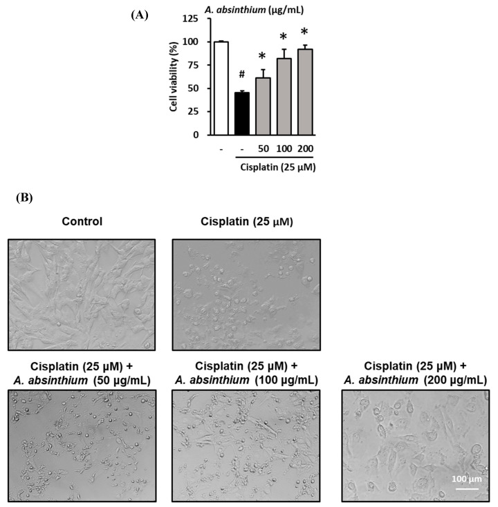
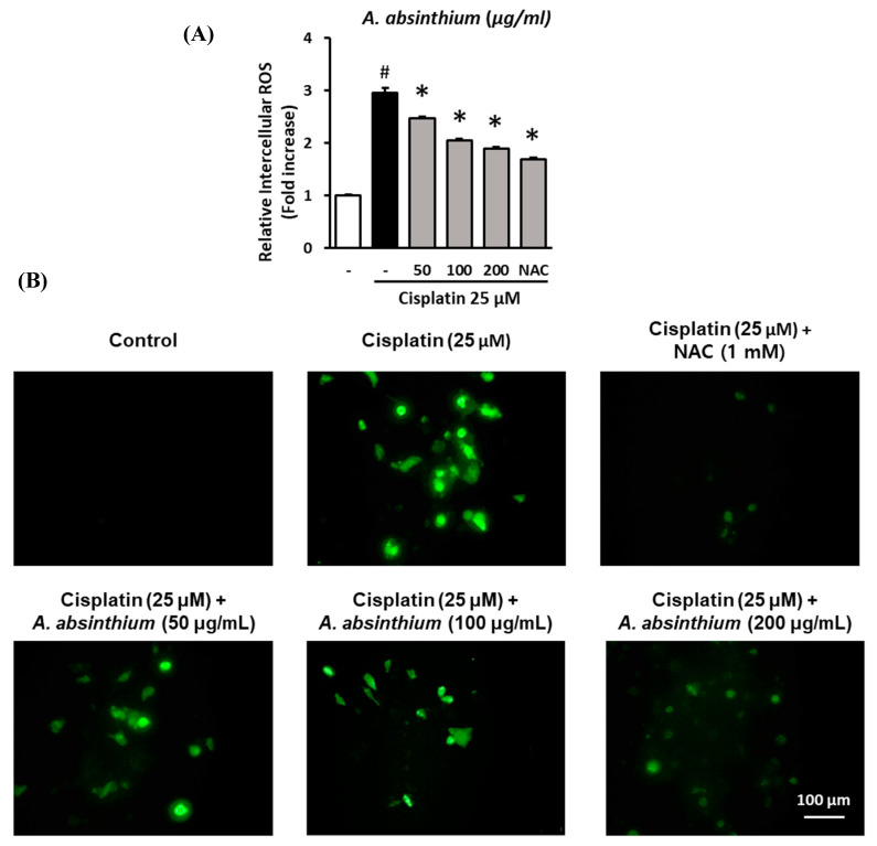
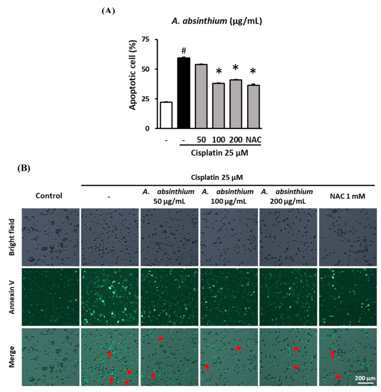
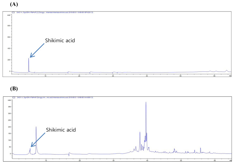
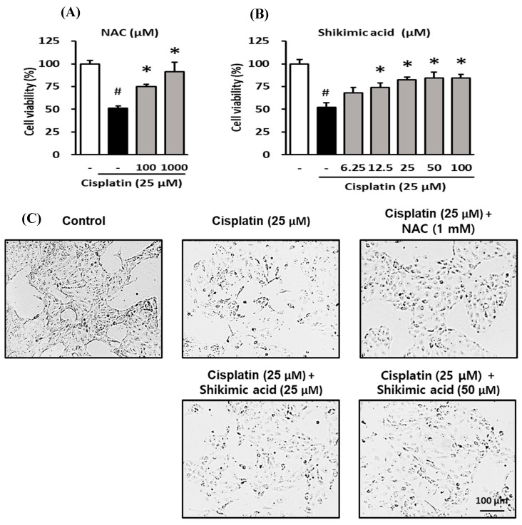
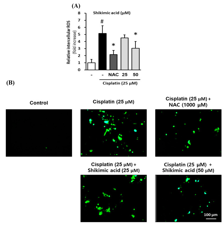
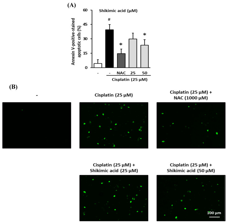
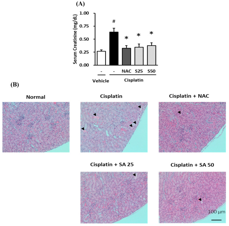
Nephrotoxicity is a serious side effect of cisplatin, which is one of the most frequently used drugs for cancer treatment. This study aimed to assess the renoprotective effect of Artemisia absinthium extract and its bioactive compound (shikimic acid) against cisplatin-induced renal injury. An in vitro assay was performed in kidney tubular epithelial cells (LLC-PK1) with 50, 100, and 200 µg/mL A. absinthium extract and 25 and 50 µM shikimic acid, and cytotoxicity was induced by 25 µM cisplatin. BALB/c mice (6 weeks old) were injected with 16 mg/kg cisplatin once and orally administered 25 and 50 mg/kg shikimic acid daily for 4 days. The results showed that the A. absinthium extract reversed the decrease in renal cell viability induced by cisplatin, whereas it decreased the reactive oxidative stress accumulation and apoptosis in LLC-PK1 cells. Shikimic acid also reversed the effect on cell viability but decreased oxidative stress and apoptosis in renal cells compared with the levels in the cisplatin-treated group. Furthermore, shikimic acid protected against kidney injury in cisplatin-treated mice by reducing serum creatinine levels. The protective effect of shikimic acid against cisplatin-mediated kidney injury was confirmed by the recovery of histological kidney injury in cisplatin-treated mice. To the best of our knowledge, this study is the first report on the nephroprotective effect of A. absinthium extract and its mechanism of action against cisplatin-induced renal injury.

Keywords: Artemisia absinthium; apoptosis; cisplatin; nephrotoxicity; shikimic acid.


Conflict of interest statement

There are no conflicts of interest.

Figures

Figure 1
Figure 1
Protective effect of A. absinthium extract against the cisplatin-induced decrease in kidney cell viability. (A) Effect of the extract on the viability of LLC-PK1 cells exposed to 25 µM cisplatin for 24 h using the Ez-Cytox cell viability assay kit. (B) The change in morphology of LLC-PK1 cells after treatment with cisplatin and extracts. Results are the mean ± SD. The difference in the mean values between groups was assessed using the Tukey method for one-way analysis of variance (ANOVA). # p < 0.05 versus the control group (first column) and * p < 0.05 versus the cisplatin-treated group (second column). SD, standard deviation.
Figure 2
Figure 2
Inhibitory effect of A. absinthium extract on cisplatin-induced ROS accumulation in LLC-PK1 cells. LLC-PK1 cells were treated with extract and NAC 1 mM exposed 25 µM cisplatin for 24 h. Next, the intracellular ROS accumulation was assessed via fluorescence using 2′,7′-dichlorodihydrofluorescein diacetate (DCFDA). Fluorescence images of the cells were captured using an inverted microscope. (A) The ratio of ROS accumulation between the treated groups and untreated group. (B) Representative images of the fluorescence signals of intercellular ROS in LLC-PK1 cells treated with various concentrations of A. absinthium extract or NAC and cisplatin (Green). ROS, reactive oxygen species; DCFDA, 2′,7′-dichlorodihydrofluorescein diacetate; NAC, N-acetyl-l-cysteine. Results are the mean ± SD. The difference in the mean values between groups was assessed using the Tukey method for one-way analysis of variance (ANOVA). # p < 0.001 versus the control group (first column) and * p < 0.001 versus the cisplatin-treated group (second column). SD, standard deviation.
Figure 3
Figure 3
Inhibitory effect of A. absinthium extract on cisplatin-induced apoptosis in kidney cells. LLC-PK1 cells were treated with A. absinthium extract and 25 µM cisplatin for 24 h and then stained with Alexa Fluor 488 annexin V conjugate. The number of apoptotic cells was determined with TaliPCApp software. Next, fluorescence images of the cells were captured using a Tali Image-based cytometer. (A) The percentage of apoptotic LLC-PK1 cells between the treated groups and untreated group. (B) Representative images of the fluorescence signal indicating apoptosis in LLC-PK1 cells treated with various concentrations of A. absinthium extract or NAC and 25 µM cisplatin for 24 h using Alexa Fluor 488 annexin V conjugate staining (Green). NAC, N-acetyl-l-cysteine. Results are the mean ± SD. The difference in the mean values between groups was assessed using the Tukey method for one-way analysis of variance (ANOVA). # p < 0.001 versus the control group (first column) and * p < 0.001 versus the cisplatin-treated group (second column). SD, standard deviation. The red arrows indicate apoptotic bodies.
Figure 4
Figure 4
HPLC chromatograms of standard shikimic acid (A) and HPLC chromatograms of A. absinthium extract (B).
Figure 5
Figure 5
Protective effect of shikimic acid against cisplatin-induced kidney cell damage. The cell viability of LLC-PK1 cell was assessed using an Ez-Cytox cell viability assay kit, and the morphological changes in the cells were assessed using an inverted microscope. (A) Effect of NAC on the viability of LLC-PK1 cells exposed to 25 µM cisplatin for 24 h. (B) Effect of shikimic acid on the viability of LLC-PK1 cells exposed to 25 µM cisplatin for 24 h. (C) Effect of NAC and shikimic acid on cell density and morphology changes in LLC-PK1 cells exposed to 25 µM cisplatin for 24 h. Results are the mean ± SD. The difference in the mean values between groups was assessed using the Tukey method for one-way analysis of variance (ANOVA). # p < 0.001 versus the control group (first column) and * p < 0.001 versus the cisplatin-treated group (second column). NAC, N-acetyl-l-cysteine; SD, standard deviation.
Figure 6
Figure 6
Inhibitory effect of shikimic acid on cisplatin-induced ROS accumulation in LLC-PK1 cells. LLC-PK1 cells were treated with 25 or 50 µM shikimic acid and 25 µM cisplatin for 24 h. Next, the intracellular ROS accumulation was assessed via fluorescence using DCFDA. Fluorescence images of the cells were captured using an inverted microscope. (A) The ratio of ROS accumulation between the treated groups and untreated group. (B) Representative images of the fluorescence signals of intercellular ROS in LLC-PK1 cells treated with various concentrations of shikimic acid or NAC and cisplatin (Green). ROS, reactive oxygen species; DCFDA, 2′,7′-dichlorodihydrofluorescen diacetate; NAC, N-acetyl-l-cysteine. Results are the mean ± SD. The difference in the mean values between groups was assessed using the Tukey method for one-way analysis of variance (ANOVA). # p < 0.001 versus the control group (first column) and * p < 0.001 versus the cisplatin-treated group (second column). SD, standard deviation.
Figure 7
Figure 7
Inhibitory effect of shikimic acid on cisplatin-induced apoptosis in kidney cells. LLC-PK1 cells were treated with 25 or 50 µM shikimic acid and 25 µM cisplatin for 24 h and then stained with Alexa Fluor 488 annexin V conjugate. The number of apoptotic cells was determined with TaliPCApp software. Next, fluorescence images of the cells were captured using a Tali Image-based cytometer. (A) The percentage of apoptotic LLC-PK1 cells between the treated groups and untreated group. (B) Representative images of the fluorescence signal indicating apoptosis in LLC-PK1 cells treated with various concentrations of shikimic acid or NAC and 25 µM cisplatin for 24 h using Alexa Fluor 488 annexin V conjugate staining (Green). Results are the mean ± SD. NAC, N-acetyl-l-cysteine. The difference in the mean values between groups was assessed using the Tukey method for one-way analysis of variance (ANOVA). # p < 0.001 versus the control group (first column) and * p < 0.001 versus the cisplatin-treated group (second column). SD, standard deviation.
Figure 8
Figure 8
Protective effect of shikimic acid against the cisplatin-induced serum creatinine increase and histological kidney injury in 6-week-old BALB/c mice. Mice were injected with physiological saline (normal) or 16 mg/kg cisplatin (cisplatin) in physiological saline before being treated with NAC (1000 mg/kg) or shikimic acid (25 mg/kg, SA25, and 50 mg/kg, SA50) (n = 5–6 mice/group) for 4 d. Serum creatinine was measured using an AceChem Creatinine kit. (A) Serum creatinine levels in mice treated with shikimic acid or NAC after being injected with cisplatin. (B) Hematoxylin & Eosin (H&E) staining of representative kidney sections of the shikimic-acid- and NAC-treated mice after being injected with cisplatin. Black arrows indicate an area of kidney injury. Results are the mean ± SEM. NAC, N-acetyl-l-cysteine; SA, shikimic acid; SEM, standard error of the mean; H&E, hematoxylin & eosin. The difference in the mean values between groups was assessed using the Tukey method for one-way analysis of variance (ANOVA). # p < 0.001 versus the control group (first column) and * p < 0.05 versus the cisplatin-treated group (second column).

Similar articles

Cited by

References

    1. Rosenberg B.H., Vancamp L., Trosko J.E., Mansour V.H. Platinum Compounds: A New Class of Potent Antitumour Agents. Nat. Cell Biol. 1969;222:385–386. doi: 10.1038/222385a0. - DOI - PubMed
    1. Rosenberg B., Vancamp L. The successful regression of large solid sarcoma 180 tumors by platinum compounds. Cancer Res. 1970;30:1799–1802. - PubMed
    1. Higby D.J., Wallace H.J., Jr., Albert D., Holland J. Diaminodichloroplatinum: A phase I study showing responses in testicular and other tumors. Cancer. 1974;33:1219–1225. doi: 10.1002/1097-0142(197405)33:5<1219::AID-CNCR2820330505>3.0.CO;2-U. - DOI - PubMed
    1. Erdlenbruch B., Pekrum A., Roth C., Grunewald R.W., Kern W., Lakomek M. Cisplatin nephrotoxicity in children after continuous 72-h and 3x1-h infusions. Pediatr. Nephrol. 2001;16:586–593. doi: 10.1007/s004670100610. - DOI - PubMed
    1. Leu L., Baribeault D. A comparison of the rates of cisplatin (cDDP)-induced nephrotoxicity associated with sodium loading or sodium loading with forced diuresis as a preventative measure. J. Oncol. Pharm. Pract. 2009;16:167–171. doi: 10.1177/1078155209346071. - DOI - PubMed

LinkOut - more resources