Review
. 2020 Dec 1;25(23):5662.
doi: 10.3390/molecules25235662.

Quinolones: Mechanism, Lethality and Their Contributions to Antibiotic Resistance

Affiliations
Review

Quinolones: Mechanism, Lethality and Their Contributions to Antibiotic Resistance

Natassja G Bush et al. Molecules. .

Abstract

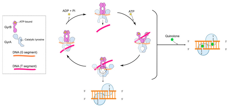
Fluoroquinolones (FQs) are arguably among the most successful antibiotics of recent times. They have enjoyed over 30 years of clinical usage and become essential tools in the armoury of clinical treatments. FQs target the bacterial enzymes DNA gyrase and DNA topoisomerase IV, where they stabilise a covalent enzyme-DNA complex in which the DNA is cleaved in both strands. This leads to cell death and turns out to be a very effective way of killing bacteria. However, resistance to FQs is increasingly problematic, and alternative compounds are urgently needed. Here, we review the mechanisms of action of FQs and discuss the potential pathways leading to cell death. We also discuss quinolone resistance and how quinolone treatment can lead to resistance to non-quinolone antibiotics.

Keywords: DNA gyrase; DNA topology; antibacterials; antibiotic resistance; fluoroquinolones; supercoiling; topoisomerases.


Conflict of interest statement

The authors declare no conflict of interest. The funders had no role in the design of this study or the decision to publish.

Figures

Figure 1
Figure 1
Chemical structures of several significant fluoroquinolones. Nalidixic acid (the first “quinolone”) is shown, along with the chloroquine by-product inspiring its synthesis. Note that nalidixic acid lacks the 4-quinolone core and instead contains a 1,8-naphthyridine nucleus.
Figure 2
Figure 2
The observed structure-activity relationships (SARs) of quinolone core substitutions. Most often R5 = H, but sparfloxacin (a discontinued 3rd-generation fluoroquinolone (FQ)) has R5 = NH2 (diagram adapted from [32], with permission).
Figure 3
Figure 3
Structures of “new” fluoroquinolones and non-fluoroquinolone topoisomerase inhibitors discussed in the text. Gepotidacin and Zoliflodacin (Novel Bacterial Topoisomerase Inhibitors) are both in phase III clinical trials. Thiophene 2: N-(2-amino-1-phenylethyl)-5-(2-chlorophenyl)-2-methylthiophene-3-carboxamide, Imidazopyrazinone t1: 7-((3~[S])-3-azanylpyrrolidin-1-yl)-.5-cyclopropyl-8-fluoranyl-imidazo (1,2-a)quinoxalin-4-one and Quinazolinedione UING5-207: 3-Amino-1-cyclopropyl-7-((3R)-3-ethyl-1-pyrrolidinyl)-6-fluoro-8-methoxy-2,4(1H,3H)-quinazolinedione.
Figure 4
Figure 4
Cartoon showing the proposed mechanism of DNA supercoiling by DNA gyrase and how quinolones interfere with this mechanism by stabilising the cleavage complex. The inset shows the GyrA (blue) and GyrB (purple) subunits. Y indicates the position of the active site tyrosine, and the star indicates the position of the ATP-binding site. The G segment (orange) binds across the GyrA dimer interface. The GyrA C-terminal domain wraps the DNA (not shown) to present the T segment (pink) in a positive node. ATP binds to the N-terminal domain of GyrB, which closes the GyrB clamp (also known as the N-gate), capturing the T segment. The G segment is transiently cleaved, the GyrB domains rotate (not shown), the DNA gate widens and the T segment is transported through the cleaved G segment. The G segment is re-ligated, and the T segment exits through the GyrA C-gate. The hydrolysis of ATP and the leaving of ADP + Pi resets the enzyme for another cycle, although the exact timing of these reactions is unknown. The black-dashed circle and lower inset show the cleavage complex. The right-hand panel shows the binding of quinolones (green spheres) in the cleavage complex.
Figure 5
Figure 5
Overview of moxifloxacin binding to the topo IV-DNA complex (Protein Data Bank (PDB): 2XKK; [55]; figure modified with permission). (a) Front-faced view of moxifloxacin (Van der Waals model, green carbons) bound within the cleavage complex of Acinetobacter baumannii topo IV ParE (purple ribbons) N-terminal domain, fused to a ParC (blue ribbons) C-terminal domain and complexed with a 34-base pair (bp) heteroduplex DNA (yellow and coral ribbons). (b) View of the same complex from above. (c) Detail of moxifloxacin (ball and stick, green carbons) partially intercalated into the DNA bases at the break sites, spaced 4-bp apart. (d) Water-metal ion bridge formed between moxifloxacin, a noncatalytic Mg2+ (grey sphere), four water molecules (red spheres) and S84 and G88 of ParC.
Figure 6
Figure 6
Model of quinolone lethality. (1) Quinolones stabilise the topoisomerase-DNA cleavage complex in which there is a double-strand break. (2) If the cleavage complex is not resolved, (3) replication and transcription cannot happen, which causes slow bacterial cell death. (4) If the topoisomerase is removed, the double-strand break is free, and if left unrepaired, (5) it leads to the fragmentation of the chromosome, which causes rapid bacterial cell death. (6) The stabilised cleavage complex, or the removal of the topoisomerase from the cleavage complex, might lead to the accumulation of reactive oxygen species (ROS) that can cause rapid bacterial cell death.
Figure 7
Figure 7
Contributions of quinolones to antibiotic resistance. Quinolones can select for quinolone resistance, which is caused by the upregulation of efflux pumps, mutations in DNA gyrase or DNA topoisomerase IV genes or plasmid-encoded resistance genes. Quinolones can also induce resistance to quinolones and non-quinolones antibiotics, presumably by the activation of a stress response that then increases the mutation, recombination or persister formation rates.

References

    1. Lesher G.Y., Froelich E.J., Gruett M.D., Bailey J.H., Brundage R.P. 1,8-Naphthyridone derivatives. A new class of chemotherapeutic agents. J. Med. Pharm. Chem. 1962;91:1063–1065. doi: 10.1021/jm01240a021. - DOI - PubMed
    1. Bisacchi G.S. Origins of the quinolone class of antibacterials: An expanded “Discovery Story”. J. Med. Chem. 2015;58:4874–4882. doi: 10.1021/jm501881c. - DOI - PubMed
    1. Gillespie S.H. The role of moxifloxacin in tuberculosis therapy. ERR. 2016;25:19–28. doi: 10.1183/16000617.0085-2015. - DOI - PMC - PubMed
    1. Oliphant C.M., Green G.M. Quinolones: A comprehensive review. Am. Fam. Physician. 2002;65:455–464. - PubMed
    1. Aldred K.J., Kerns R.J., Osheroff N. Mechanism of quinolone action and resistance. Biochemistry. 2014;53:1565–1574. doi: 10.1021/bi5000564. - DOI - PMC - PubMed

MeSH terms