Review
. 2020 Dec 1;21(23):9170.
doi: 10.3390/ijms21239170.

Targeting HGF/c-MET Axis in Pancreatic Cancer

Affiliations
Review

Targeting HGF/c-MET Axis in Pancreatic Cancer

Srinivasa P Pothula et al. Int J Mol Sci. .

Abstract

Pancreatic cancer (pancreatic ductal adenocarcinoma (PDAC/PC)) has been an aggressive disease that is associated with early metastases. It is characterized by dense and collagenous desmoplasia/stroma, predominantly produced by pancreatic stellate cells (PSCs). PSCs interact with cancer cells as well as other stromal cells, facilitating disease progression. A candidate growth factor pathway that may mediate this interaction is the hepatocyte growth factor (HGF)/c-MET pathway. HGF is produced by PSCs and its receptor c-MET is expressed on pancreatic cancer cells and endothelial cells. The current review discusses the role of the MET/HGF axis in tumour progression and dissemination of pancreatic cancer. Therapeutic approaches that were developed targeting either the ligand (HGF) or the receptor (c-MET) have not been shown to translate well into clinical settings. We discuss a two-pronged approach of targeting both the components of this pathway to interrupt the stromal-tumour interactions, which may represent a potential therapeutic strategy to improve outcomes in PC.

Keywords: HGF-c-MET; Pancreatic Cancer; Stromal-tumour interactions.


Conflict of interest statement

The authors declare no conflict of interest.

Figures

Figure 1
Figure 1
Representative photomicrograph of haematoxylin & eosin stained human pancreatic cancer section showing malignant elements (duct-like and tubular structures-indicated by asterisks) embedded in a highly fibrotic stromal reaction (indicated by arrows). Reprinted with permission from Elsevier [4].
Figure 2
Figure 2
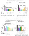
Effects of HGF/c-MET inhibition with and without gemcitabine on pancreatic cancer progression in orthotopic models of early and advanced pancreatic cancer. Pancreatic tumours were produced by injection of a mixture of human pancreatic cancer cells + human pancreatic stellate cells into the pancreas of Balb/c nude mice. For the early pancreatic cancer model, treatment was commenced one week after cell implantation, while for the advanced PC model treatment was commenced 4 weeks after cell implantation. In early pancreatic cancer, while single and dual combinations of the HGF neutralising antibody (HGF Inhib), c-MET inhibitor (cMET Inhib) and gemcitabine (Gem) reduced tumour volume compared to control (untreated) mice (IgG), the greatest reduction in tumour volume was observed in mice treated with the triple therapy. In advanced PC, the effects of single or dual agents on tumour volumes were variable, however, the greatest reduction in tumour volume was again observed in mice treated with triple therapy. Importantly, triple therapy had a striking effect on metastasis, with virtual elimination in early pancreatic cancer (only one liver nodule observed in one mouse) and complete absence of metastasis in advanced pancreatic cancer [10,53]).
Figure 3
Figure 3
Schematic depiction of the possible role of HGF/c-MET pathway in pancreatic cancer. HGF secreted by PSCs in a precursor form is activated by proteolytic cleavage, mediated by proteases such as urokinase plasminogen activator (uPA). Upon binding of HGF to its receptor c-MET, downstream intracellular signalling cascades are activated which regulate cancer cell functions that influence tumour progression. This binding also increases production of uPA by cancer cells that further activates HGF from PSCs, thus forming a feed-forward loop. Adapted from Pothula et al. [53].

References

    1. Siegel R.L., Miller K.D., Jemal A. Cancer statistics, 2020. CA Cancer J. Clin. 2020;70:7–30. doi: 10.3322/caac.21590. - DOI - PubMed
    1. Rahib L., Smith B.D., Aizenberg R., Rosenzweig A.B., Fleshman J.M., Matrisian L.M. Projecting cancer incidence and deaths to 2030: The unexpected burden of thyroid, liver, and pancreas cancers in the United States. Cancer Res. 2014;74:2913–2921. doi: 10.1158/0008-5472.CAN-14-0155. - DOI - PubMed
    1. Feig C., Gopinathan A., Neesse A., Chan D.S., Cook N., Tuveson D.A. The Pancreas Cancer Microenvironment. Clin. Cancer Res. 2012;18:4266–4276. doi: 10.1158/1078-0432.CCR-11-3114. - DOI - PMC - PubMed
    1. Apte M.V., Xu Z., Pothula S., Goldstein D., Pirola R., Wilson J. Pancreatic cancer: The microenvironment needs attention too! Pancreatology. 2015;15(Suppl. S4):S32–S38. doi: 10.1016/j.pan.2015.02.013. - DOI - PubMed
    1. Apte M.V., Park S., Phillips P.A., Santucci N., Goldstein D., Kumar R.K., Ramm G.A., Buchler M., Friess H., McCarroll J.A., et al. Desmoplastic reaction in pancreatic cancer: Role of pancreatic stellate cells. Pancreas. 2004;29:179–187. doi: 10.1097/00006676-200410000-00002. - DOI - PubMed

MeSH terms