. 2020 Dec 1;21(23):9171.
doi: 10.3390/ijms21239171.

The Impact of Human Lipoaspirate and Adipose Tissue-Derived Stem Cells Contact Culture on Breast Cancer Cells: Implications in Breast Reconstruction

Affiliations

The Impact of Human Lipoaspirate and Adipose Tissue-Derived Stem Cells Contact Culture on Breast Cancer Cells: Implications in Breast Reconstruction

Asim Ejaz et al. Int J Mol Sci. .

Abstract

Background: Autologous fat transfer in the form of lipoaspirates for the reconstruction of the breast after breast cancer surgery is a commonly used procedure in plastic surgery. However, concerns regarding the oncologic risk of nutrient-rich fat tissue are widely debated. Previous studies have primarily focused on studying the interaction between adipose-derived stem cells (ASCs) and breast cancer cells.

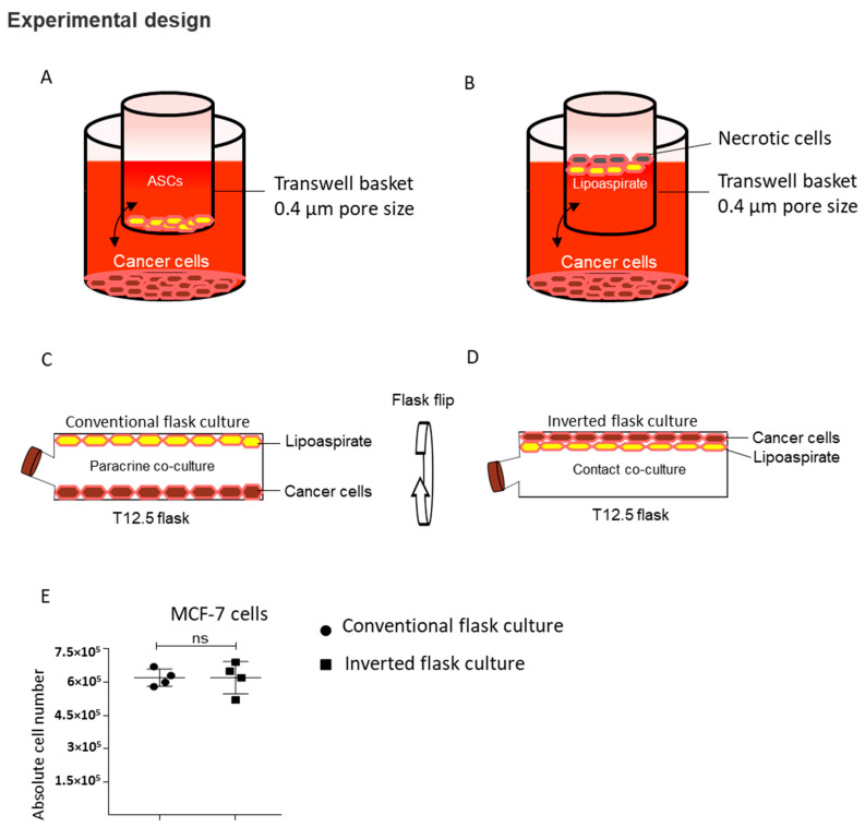
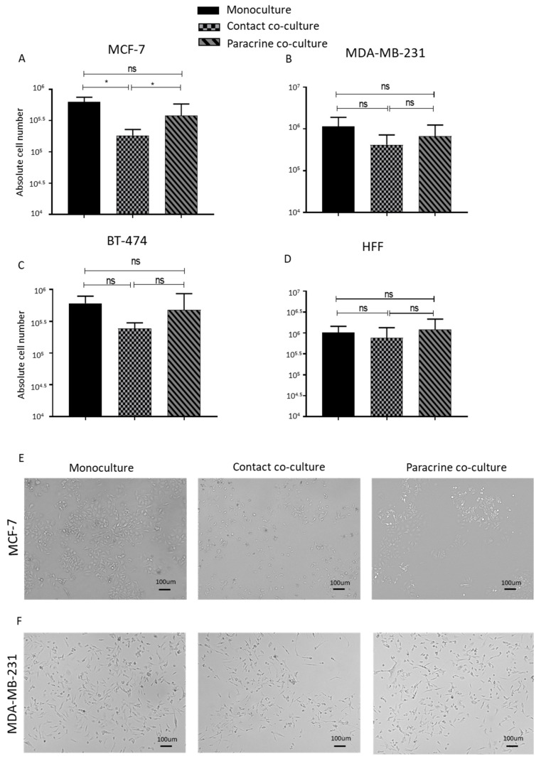
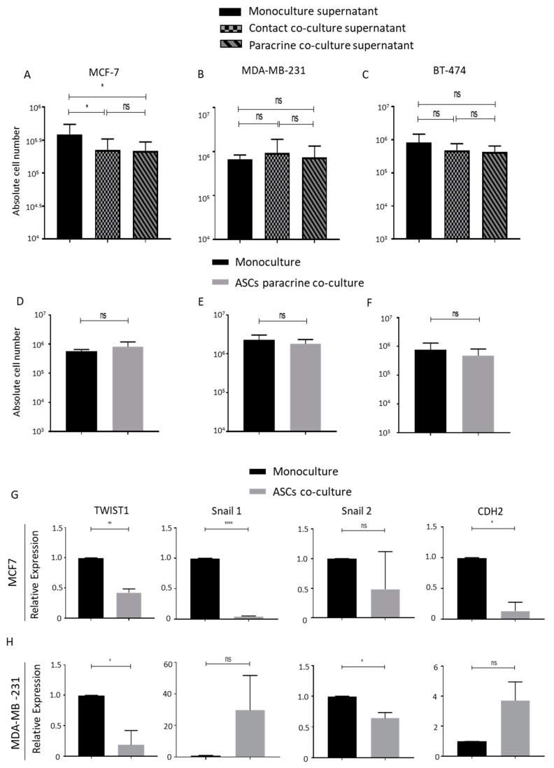
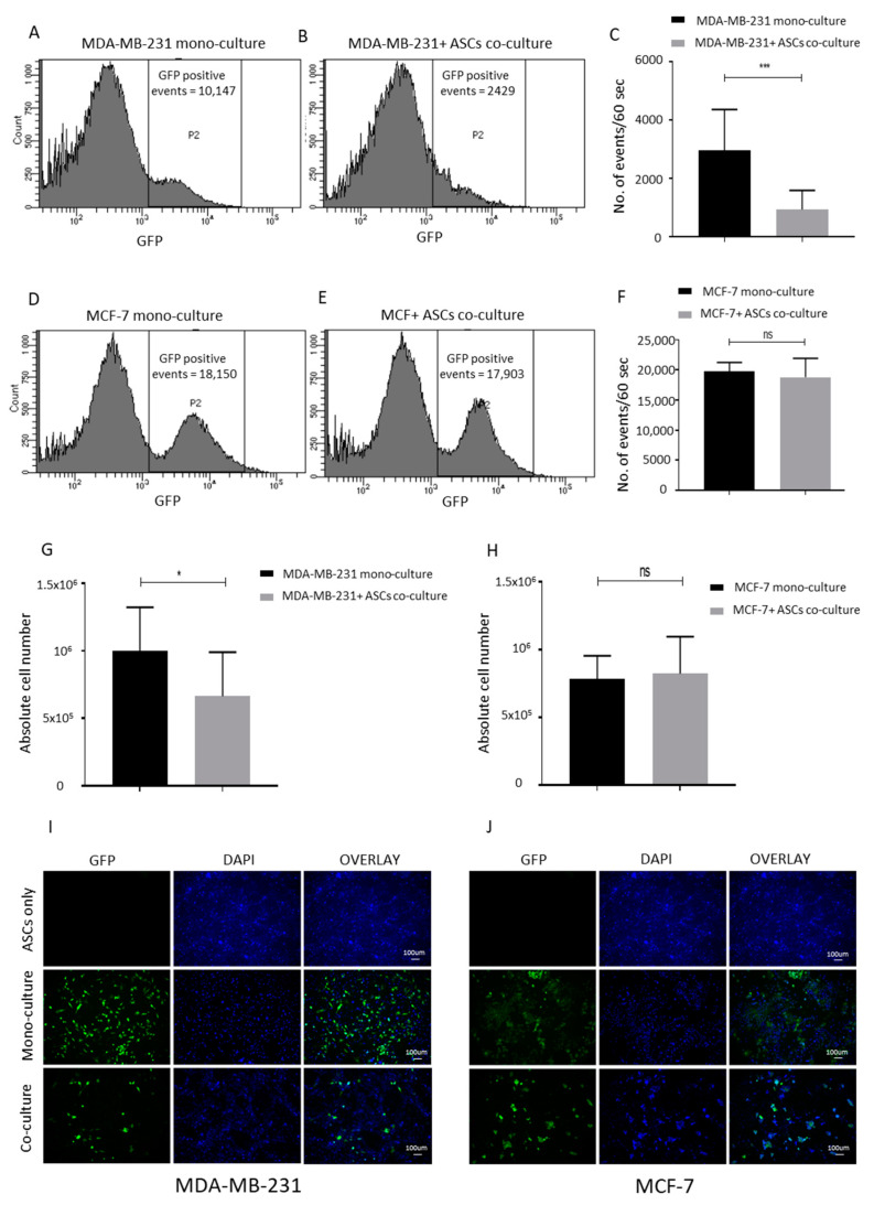
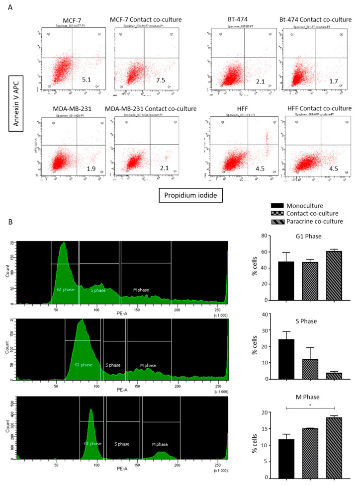
Methods: In this study, we performed a comprehensive analysis of the paracrine- and contact-based interactions between lipoaspirates, ASCs and breast cancer cell lines. An inverted flask culture method was used to study the contact-based interaction between lipoaspirates and breast cancer cells, while GFP-expressing breast cancer cell lines were generated to study the cell-cell contact interaction with ASCs. Three different human breast cancer cell lines, MCF-7, MDA-MB-231 and BT-474, were studied. We analyzed the impact of these interactions on the proliferation, cell cycle and epithelial-to-mesenchymal (EMT) transition of the breast cancer cells.

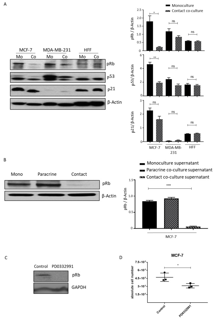
Results: Our results revealed that both lipoaspirates and ASCs do not increase the proliferation rate of the breast cancer cells either through paracrine- or contact-dependent interactions. We observed that lipoaspirates selectively inhibit the proliferation of MCF-7 cells in contact co-culture, driven by the retinoblastoma (Rb) protein activity mediating cell cycle arrest. Additionally, ASCs inhibited MDA-MB-231 breast cancer cell proliferation in cell-cell contact-dependent interactions. Quantitative real-time PCR revealed no significant increase in the EMT-related genes in breast cancer cells upon co-culture with ASCs.

Conclusion: In conclusion, this study provides evidence of the non-oncogenic character of lipoaspirates and supports the safety of clinical fat grafting in breast reconstruction after oncological surgical procedures. In vivo studies in appropriate animal models and long-term post-operative clinical data from patients are essential to reach the final safety recommendations.

Keywords: adipose-derived stem cells; breast cancer cells; cell cycle and proliferation; lipoaspirates.


Conflict of interest statement

The authors indicated no potential conflict of interest.

Figures

Figure 1
Figure 1
Contact and paracrine co-culture experimental design. (A) Paracrine culture setup of cancer cells with adipose-derived stem cells (ASCs) using 6-well transwell plates. (B) Paracrine culture setup of cancer cells with lipoaspirates. (C,D) Modified paracrine (C) and contact (D) co-culture of cancer cells with lipoaspirate using T12.5 cm2 flask. (E) Comparison of MCF-7 cells proliferation in conventional and inverted flask culture (n = 4). ns = non-significant.
Figure 2
Figure 2
Contact and paracrine co-culture of lipoaspirate do not promote breast cancer cells’ proliferation. (AD) Absolute cell count obtained using Neubauer counting chamber after 4 days of either contact or paracrine co-culture of lipoaspirates with MCF-7 (A), MDA-MB-231 (B), BT-474 (C), or human foreskin fibroblast (HFF) (D). Graphs are representative of the results obtained using lipoaspirate from 3 different donors. p value < 0.05 = *, ns = non-significant. (E,F) Bright field microscope images of contact and paracrine co-culture of MCF-7 and MDA-MB-231 cells with lipoaspirates.
Figure 3
Figure 3
Transwell paracrine culture of ASCs does not promote breast cancer cells proliferation. (AC) Absolute cell count obtained using Neubauer counting chamber after 4 days of culture of either MCF-7 (A), MDA-MB-231 (B) or BT-474 (C), in cell culture supernatant collected from either monocultured, contact-co-cultured or paracrine-cultured breast cancer cells with lipoaspirates. Graphs are representative of 3 independent experiments. (DF) Representative graph of cell count of MCF-7 (D), MDA-MB-231 (E) and BT-474 (F) cell upon transwell co-culture with ASCs. ASCs from 3 different donors were employed. (G,H) Relative EMT genes expression of monocultured or co-cultured MCF-7 (G) and MDA-MB-231 (H) was analyzed by quantitative real-time PCR. p value < 0.05 = *, p < 0.01 = **, p < 0.0001 = ****, ns = non-significant.
Figure 4
Figure 4
ASCs contact culture inhibits the proliferation of MDA-MB-231 cells. (AC) Contact co-culture of MDA-MB-231 cells with ASCs. MDA-MB-231 expressing GFP were seeded either alone (A) or on confluent ASCs (B) for 4 days. The numbers of GFP-positive events per minute were counted 4 days post culture by using flow cytometry and plotted (C). (DF) Contact co-culture of MCF-7 cells with ASCs. MCF-7 cells expressing GFP were seeded either alone (D) or on confluent ASCs (E) for 4 days. The numbers of GFP positive events per minute were counted 4 days post culture by using flow cytometry, and plotted (F). The graphs are representative of 3 independent experiments using ASCs from different donors. (G,H) Absolute number of cells counted by Neubauer chamber from the experimental settings (A,B) shown in (G) and from (D,E) as reflected in (H). (I,J) Fluorescent microscope images of MDA-MB-231/ASCs co-culture (I) and MCF-7/ASCs co-culture (J) are shown. GFP-positive cells are green while nuclear stain DAPI stain blue. p value < 0.05 = *, p < 0.001 = ***, ns = non-significant.
Figure 5
Figure 5
Lipoaspirate contact culture results in cell cycle arrest in MCF-7 cells. (A) Effect of lipo-aspirate contact co-culture on breast cancer cell survival. AnnexinV/Propidium Iodide staining was employed to estimate the cell death of breast cancer cells upon co-culture. (B) Cell cycle analyses of paracrine and contact-co-cultured MCF-7 cells with lipo-aspirates. Graphs show the percentages of cells in different phases of cells upon different culture conditions. p value < 0.05 = *.
Figure 6
Figure 6
Contact lipoaspirate culture inhibits MCF-7 proliferation via the retinoblastoma-mediated pathway. (A) Cell lysates from MCF-7, MDA-MB-231 or HFF contact-co-cultured with lipo-aspirates were collected in Ripa lysate buffer. The phosphorylation of retinoblastoma protein (Rb) and the expression of p53 and p21 were analyzed by western blotting using specific antibodies. β-Actin was employed as the loading control. Fold changes in densitometric band intensities, acquired by image J and normalized to β-Actin, were compared and plotted. (B) Cell lysates from MCF-7 cells cultured in media from either monocultured, paracrine-cultured or contact-cultured MCF-cells with lipo-aspirates were collected in Ripa buffer and blotted for phosphorylated retinoblastoma protein (Rb). β-Actin was employed as the loading control. (C,D) MCF-7 cells were incubated with 10µM PD0332991 and cell lysates were blotted for Rb protein phosphorylation (C) and counted at day 4 post seeding (D). p value < 0.05 = *, p < 0.01 = **, p < 0.001 = ***, ns = non-significant.

References

    1. Krastev T., van Turnhout A., Vriens E., Smits L., van der Hulst R. Long-term follow-up of autologous fat transfer vs conventional breast reconstruction and association with cancer relapse in patients with breast cancer. JAMA Surg. 2019;154:56–63. doi: 10.1001/jamasurg.2018.3744. - DOI - PMC - PubMed
    1. Koellensperger E., Bonnert L.C., Zoernig I., Marmé F., Sandmann S., Germann G., Gramley F., Leimer U. The impact of human adipose tissue-derived stem cells on breast cancer cells: Implications for cell-assisted lipotransfers in breast reconstruction. Stem Cell Res. Ther. 2017;8:121. doi: 10.1186/s13287-017-0579-1. - DOI - PMC - PubMed
    1. Coleman S.R. Structural fat grafting. Aesthet. Surg. J. 1998;18:386–388. doi: 10.1016/S1090-820X(98)70098-6. - DOI - PubMed
    1. Tsuji W., Valentin J.E., Marra K.G., Donnenberg A.D., Donnenberg V.S., Rubin J.P. An animal model of local breast cancer recurrence in the setting of autologous fat grafting for breast reconstruction. Stem Cells Transl. Med. 2018;7:125–134. doi: 10.1002/sctm.17-0062. - DOI - PMC - PubMed
    1. Krastev T.K., Alshaikh G.A., Hommes J., Piatkowski A., van der Hulst R.R. Efficacy of autologous fat transfer for the correction of contour deformities in the breast: A systematic review and meta-analysis. J. Plast. Reconstr. Aesthet. Surg. 2018;71:1392–1409. doi: 10.1016/j.bjps.2018.05.021. - DOI - PubMed