. 2020 Dec 1;21(23):9176.
doi: 10.3390/ijms21239176.

RB Regulates DNA Double Strand Break Repair Pathway Choice by Mediating CtIP Dependent End Resection

Affiliations

RB Regulates DNA Double Strand Break Repair Pathway Choice by Mediating CtIP Dependent End Resection

Yuning Jiang et al. Int J Mol Sci. .

Abstract

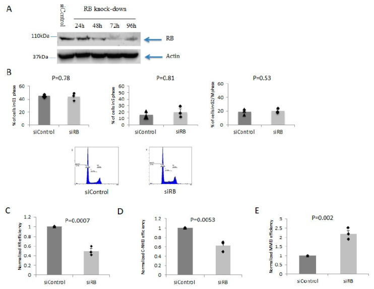
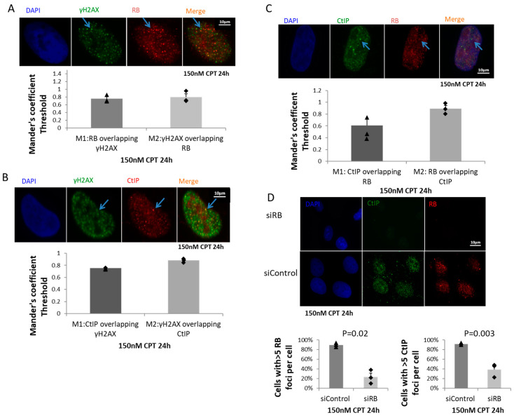
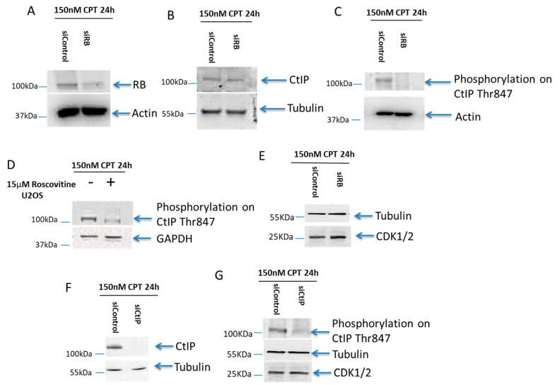
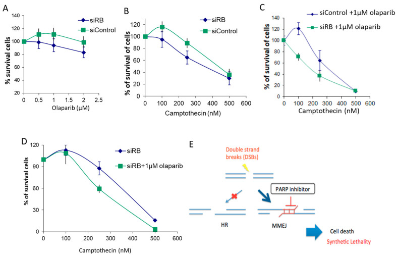
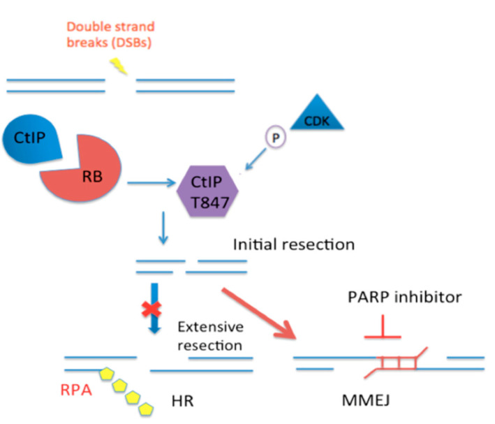
Inactivation of the retinoblastoma tumor suppressor gene (RB1) leads to genome instability, and can be detected in retinoblastoma and other cancers. One damaging effect is causing DNA double strand breaks (DSB), which, however, can be repaired by homologous recombination (HR), classical non-homologous end joining (C-NHEJ), and micro-homology mediated end joining (MMEJ). We aimed to study the mechanistic roles of RB in regulating multiple DSB repair pathways. Here we show that HR and C-NHEJ are decreased, but MMEJ is elevated in RB-depleted cells. After inducing DSB by camptothecin, RB co-localizes with CtIP, which regulates DSB end resection. RB depletion leads to less RPA and native BrdU foci, which implies less end resection. In RB-depleted cells, less CtIP foci, and a lack of phosphorylation on CtIP Thr847, are observed. According to the synthetic lethality principle, based on the altered DSB repair pathway choice, after inducing DSBs by camptothecin, RB depleted cells are more sensitive to co-treatment with camptothecin and MMEJ blocker poly-ADP ribose polymerase 1 (PARP1) inhibitor. We propose a model whereby RB can regulate DSB repair pathway choice by mediating the CtIP dependent DNA end resection. The use of PARP1 inhibitor could potentially improve treatment outcomes for RB-deficient cancers.

Keywords: CtIP; RB; classical non-homologous end joining; homologous recombination; micro-homology mediated end joining; resection.


Conflict of interest statement

The authors declare no conflict of interest.

Figures

Figure 1
Figure 1
RB knocked-down cells repair double strand breaks (DSBs) mainly by the micro-homology mediated end joining (MMEJ) pathway. (A): RB was knocked down by siRNA treatment. (B): Cell cycle distribution in both RB knocked-down cells and the control treated cells. Representative cell cycle profiles are shown from one of the triplicate experiments. (C): Homologous recombination (HR) efficiency was measured by the DRGFP reporter assay. (D): C-NHEJ efficiency was measured by the EJ5GFP reporter assay. (E): MMEJ efficiency was measured by the EJ2GFP reporter assay. All experiments were repeated three times. Bar charts show the mean values from three experiments. Error bars show the standard error of the mean. Unpaired T-test was used for statistical analysis.
Figure 2
Figure 2
RB co-localized with CtIP at DSBs and regulated CtIP foci formation. (A): RB and γH2AX were co-stained by immunofluorescence after treating cells with 150 nM CPT for 24 h. Scale bar is 10 μm. Co-localization of RB and γH2AX was quantified by ImageJ software. In total, 115 cells were counted. Manders’ coefficients showed the percentages of co-localization of RB and γH2AX. Experiments were repeated three times. Bar chart shows the mean values from three experiments. Error bars show the standard error of the mean. Blue arrows indicate an example of RB and γH2AX co-localization. (B): CtIP and γH2AX were co-stained by immunofluorescence after treating cells with 150 nM CPT for 24 h. Scale bar is 10 μm. Co-localization of CtIP and γH2AX was quantified by ImageJ software. In total 183 cells were counted. Manders’ coefficients showed the percentages of co-localization of CtIP and γH2AX. Experiments were repeated three times. Bar chart shows the mean values from three experiments. Error bars show the standard error of the mean. Blue arrows indicate an example of CtIP and γH2AX co-localization. (C): RB and CtIP were co-stained by immunofluorescence after treating cells with 150 nM CPT for 24 h. Scale bar is 10 μm. Co-localization of RB and CtIP was quantified by ImageJ software. In total 102 cells were counted. Manders’ coefficients showed the percentages of co-localization of RB and CtIP. Experiments were repeated three times. Bar chart shows the mean values from three experiments. Error bars show the standard error of the mean. Blue arrows indicate an example of RB and CtIP co-localization. (D): RB and CtIP were co-stained in both RB knocked-down cells and control treated cells. Experiments were repeated three times. In total 102 cells in the siControl, and 138 cells in the siRB, groups were counted. Bar charts show the mean value from three experiments. Error bars show the standard error of the mean. An unpaired T-test was used for statistical analysis.
Figure 3
Figure 3
RB regulated CDK2-mediated CtIP phosphorylation on Thr847. (A): RB was knocked down in cells treated with 150nM CPT. Actin was used as the loading control (B): Equal expression levels of CtIP were found in both RB knocked-down cells and control treated cells treated with 150nM CPT for 24 h. Tubulin was used as the loading control (C): A lack of phosphorylation on CtIP T847 was observed in RB knocked-down cells treated with 150nM CPT for 24 h. Actin was used as the loading control (D): Less phosphorylation on CtIP T847 was observed in U2OS cells treated with 15 μM CDK2 inhibitor, roscovitine, and 150nM CPT for 24 h. GAPDH was used as the loading control. (E): Equal expression level of CDK1/2 in both RB knocked-down cells and control treated cells treated with 150nM CPT for 24 h. Tubulin was used as the loading control. (F): CtIP was knocked down in cells treated with 150nM CPT. Tubulin was used as the loading control. (G): A lack of phosphorylation on CtIP Thr847 was detected in CtIP knocked-down cells treated with 150nM CPT for 24 h. An equal expression level of CDK1/2 was detected in CtIP knocked-down cells and control treated cells. Tubulin was used as the loading control.
Figure 4
Figure 4
RB knocked-down cells were hypersensitive to the combined treatment of poly-ADP ribose polymerase 1 (PARP1) inhibitor and camptothecin. (A): Cell survival was quantified by MTT assay in RB knocked-down cells with different dosages of olaparib for 3 days. (B): Cell survival was quantified by MTT assay in RB knocked-down and control cells treated with different dosages of CPT for 3 days. (C): Cell survival was quantified by MTT assay in RB knocked-down and control cells treated with different dosages of CPT with 1 μM olaparib for 3 days. (D): Cell survival was quantified by MTT assay in RB knocked-down cells with different dosages of CPT, with or without 1 μM olaparib, for 3 days. Mean values from three experiments are shown. Error bars show the standard error of the mean. Experiments were repeated three times. (E): RB-deficient cells are HR deficient and depend on MMEJ to repair DSB-repair. After DSB induction by CPT, PARP1 inhibitor could effectively block MMEJ, and led to enhanced cell death. The light red arrow indicates the HR pathway is inhibited in RB-deficient cells. The dark blue arrow indicates the MMEJ pathway is preferred in RB-deficient cells. The light blue arrow shows the synthetic lethality in RB-deficient cells treated with PARP inhibitor.
Figure 5
Figure 5
Proposed mechanism of RB in regulating CtIP mediated end resection for DSB-repair pathway choice. PARP1 inhibitor specifically blocking MMEJ may sensitize RB-deficient cells to co-treatment with PARP inhibitor and camptothecin. The red cross indicates inhibition of the extensive resection. The red arrow shows the initial resection would lead to MMEJ.

Similar articles

Cited by

References

    1. Harbour J.W., Dean D.C. Rb function in cell-cycle regulation and apoptosis. Nat. Cell Biol. 2000;2:E65–E67. doi: 10.1038/35008695. - DOI - PubMed
    1. Stevaux O., Dyson N.J. A revised picture of the E2F transcriptional network and RB function. Curr. Opin. Cell Biol. 2002;14:684–691. doi: 10.1016/S0955-0674(02)00388-5. - DOI - PubMed
    1. Calo E., Quintero-Estades J.A., Danielian P.S., Nedelcu S., Berman S.D., Lees J.A. Rb regulates fate choice and lineage commitment in vivo. Nat. Cell Biol. 2010;466:1110–1114. doi: 10.1038/nature09264. - DOI - PMC - PubMed
    1. Nicolay B.N., Danielian P.S., Kottakis F., Lapek J.D., Jr., Sanidas I., Miles W.O., Dehnad M., Tschöp K., Gierut J.J., Manning A.L., et al. Proteomic analysis of pRb loss highlights a signature of decreased mitochondrial oxidative phosphorylation. Genes Dev. 2015;29:1875–1889. doi: 10.1101/gad.264127.115. - DOI - PMC - PubMed
    1. Nicolay B.N., Gameiro P.A., Tschöp K., Korenjak M., Heilmann A.M., Asara J.M., Stephanopoulos G., Iliopoulos O., Dyson N.J. Loss of RBF1 changes glutamine catabolism. Genes Dev. 2013;27:182–196. doi: 10.1101/gad.206227.112. - DOI - PMC - PubMed

MeSH terms

Substances

LinkOut - more resources