. 2020 Dec 3;10(1):21191.
doi: 10.1038/s41598-020-78136-1.

Local environment effects on charged mutations for developing aggregation-resistant monoclonal antibodies

Affiliations

Local environment effects on charged mutations for developing aggregation-resistant monoclonal antibodies

Jihyeon Lee et al. Sci Rep. .

Abstract

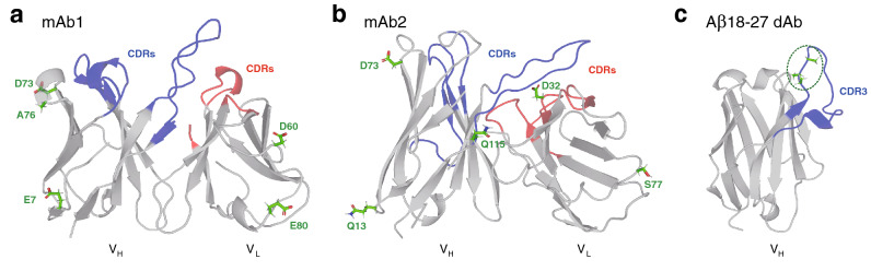
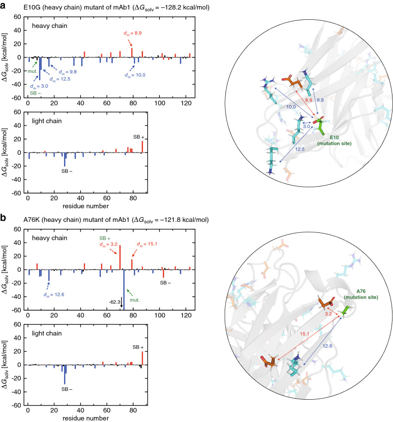
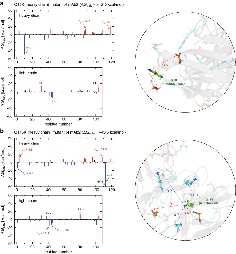
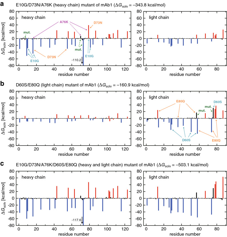
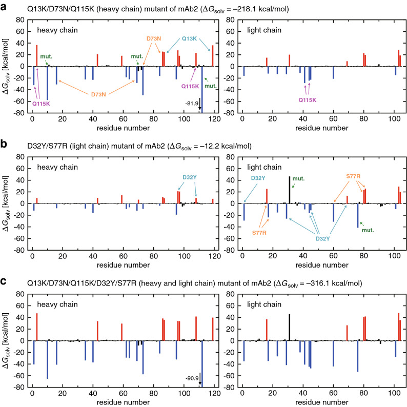
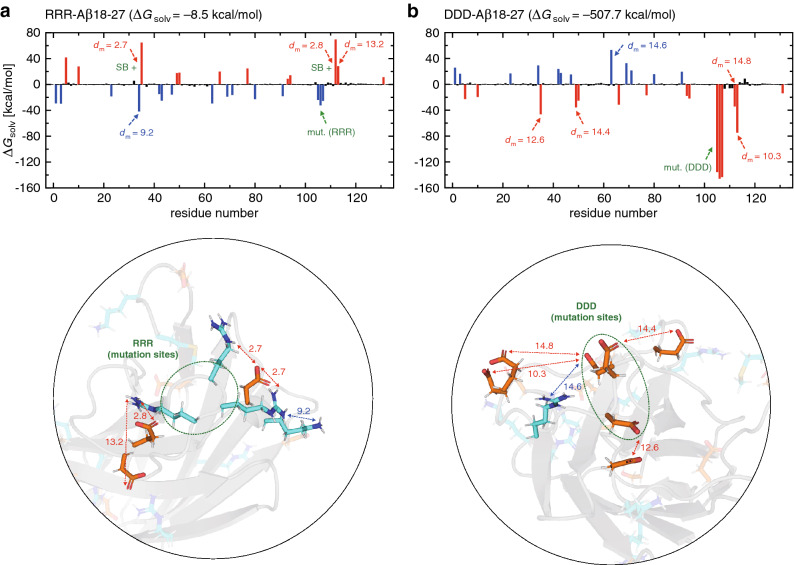
Protein aggregation is a major concern in biotherapeutic applications of monoclonal antibodies. Introducing charged mutations is among the promising strategies to improve aggregation resistance. However, the impact of such mutations on solubilizing activity depends largely on the inserting location, whose mechanism is still not well understood. Here, we address this issue from a solvation viewpoint, and this is done by analyzing how the change in solvation free energy upon charged mutation is composed of individual contributions from constituent residues. To this end, we perform molecular dynamics simulations for a number of antibody mutants and carry out the residue-wise decomposition of the solvation free energy. We find that, in addition to the previously identified "global" principle emphasizing the key role played by the protein total net charge, a local net charge within [Formula: see text]15 Å from the mutation site exerts significant effects. For example, when the net charge of an antibody is positive, the global principle states that introducing a positively charged mutation will lead to more favorable solvation. Our finding further adds that an even more optimal mutation can be done at the site around which more positively charged residues and fewer negatively charged residues are present. Such a "local" design principle accounts for the location dependence of charged mutations, and will be useful in producing aggregation-resistant antibodies.


Conflict of interest statement

The authors declare no competing interests.

Figures

Figure 1
Figure 1
(a, b) Structures of the Fv fragments comprising variable heavy (VH) and light (VL) domains of (a) the wild-type mAb1 (PDB entry 3RU8) and (b) the wild-type mAb2 (modeled based on PDB entry 1DFB). The mutation sites studied in the present work are indicated by stick representations and green labels. Heavy and light complementarity-determining regions (CDRs) are colored blue and red, respectively. (c) Structure of the single-domain (VH) antibody Aβ18-27 dAb (modeled based on PDB entry 3B9V). In the “wild type” dAb, three Ala residues (stick representations colored green) are substituted to the mutation sites (enclosed by a green dashed oval) located at the N-terminus of the third CDR (CDR3). PyMOL version 1.8.2 (https://pymol.org) was used to generate protein figures.
Figure 2
Figure 2
(a, b) Residue-wise decomposition of ΔGsolv for (a) E10G and (b) A76K mutants of mAb1 (blue, red and black colors refer to positively charged, negatively charged and neutral residues, respectively). The mutation site is indicated by the green arrow. dm (in Å) denotes the distance to the mutation site as illustrated in the inset (green stick representation, mutation site; cyan and orange representations, positively and negatively charged residues, respectively). Residues involved in the formation/breaking of salt-bridges upon mutation are represented by SB +/- (green if the mutation site is involved, and black otherwise). PyMOL version 1.8.2 (https://pymol.org) was used to generate protein figures.
Figure 3
Figure 3
(a, b) Residue-wise decomposition of ΔGsolv for (a) Q13K and (b) Q115K mutants of mAb2 (blue, red and black colors refer to positively charged, negatively charged and neutral residues, respectively). The mutation site is indicated by the green arrow. dm (in Å) denotes the distance to the mutation site as illustrated in the inset (green stick representation, mutation site; cyan and orange representations, positively and negatively charged residues, respectively). Residues involved in the formation/breaking of salt-bridges upon mutation are represented by SB +/- (green if the mutation site is involved, and black otherwise). PyMOL version 1.8.2 (https://pymol.org) was used to generate protein figures.
Figure 4
Figure 4
(ac) Residue-wise decomposition of ΔGsolv for (a) E10G/D73N/A76K, (b) D60S/E80Q, and (c) E10G/D73N/A76K/D60S/E80Q mutants of mAb1 (blue, red and black colors refer to positively charged, negatively charged and neutral residues, respectively). The mutation sites are indicated by the green arrows. Charged residues that exhibit pronounced variations in individual single-point mutations (taken from Fig. 2 and Supplementary Fig. S1) are represented by the solid arrows labeled with respective mutations. When those charged residues are deleted upon multipoint mutations, the solid arrows are replaced by the dashed ones.
Figure 5
Figure 5
(ac) Residue-wise decomposition of ΔGsolv for (a) Q13K/D73N/Q115K, (b) D32Y/S77R, and (c) Q13K/D73N/Q115K/D32Y/S77R mutants of mAb2 (blue, red and black colors refer to positively charged, negatively charged and neutral residues, respectively). The mutation sites are indicated by the green arrows. Charged residues that exhibit pronounced variations in individual single-point mutations (taken from Fig. 3 and Supplementary Fig. S2) are represented by the solid arrows labeled with respective mutations.
Figure 6
Figure 6
(a, b) Residue-wise decomposition of ΔGsolv for (a) RRR-Aβ18-27 and (b) DDD-Aβ18-27 mutants of Aβ18-27 dAb (blue, red and black colors refer to positively charged, negatively charged and neutral residues, respectively). The mutation sites are indicated by the green arrow. dm (in Å) denotes the distance to the mutation sites as illustrated in the inset (green dashed oval, mutation sites; cyan and orange stick representations, positively and negatively charged residues, respectively). Residues involved in the formation/breaking of salt-bridges upon mutation are represented by SB +/- (green if the mutation sites are involved, and black otherwise). PyMOL version 1.8.2 (https://pymol.org) was used to generate protein figures.

Similar articles

References

    1. Nelson AL, Dhimolea E, Reichert JM. Development trends for human monoclonal antibody therapeutics. Nat. Rev. Drug Discov. 2010;9:767–774. doi: 10.1038/nrd3229. - DOI - PubMed
    1. Sliwkowski MX, Mellman I. Antibody therapeutics in cancer. Science. 2013;341:1192–1198. doi: 10.1126/science.1241145. - DOI - PubMed
    1. Carter PJ, Lazar GA. Next generation antibody drugs: Pursuit of the high-hanging fruit. Nat. Rev. Drug Discov. 2018;17:197–223. doi: 10.1038/nrd.2017.227. - DOI - PubMed
    1. Lowe D, et al. Aggregation, stability, and formulation of human antibody therapeutics. Adv. Protein Chem. Struct. Biol. 2011;84:41–61. doi: 10.1016/B978-0-12-386483-3.00004-5. - DOI - PubMed
    1. Li W, et al. Antibody aggregation: Insights from sequence and structure. Antibodies. 2016;5:19. doi: 10.3390/antib5030019. - DOI - PMC - PubMed

Publication types