. 2020 Dec 3;10(1):21135.
doi: 10.1038/s41598-020-78141-4.

Mucin-2 knockout is a model of intercellular junction defects, mitochondrial damage and ATP depletion in the intestinal epithelium

Affiliations

Mucin-2 knockout is a model of intercellular junction defects, mitochondrial damage and ATP depletion in the intestinal epithelium

Mariya A Borisova et al. Sci Rep. .

Abstract

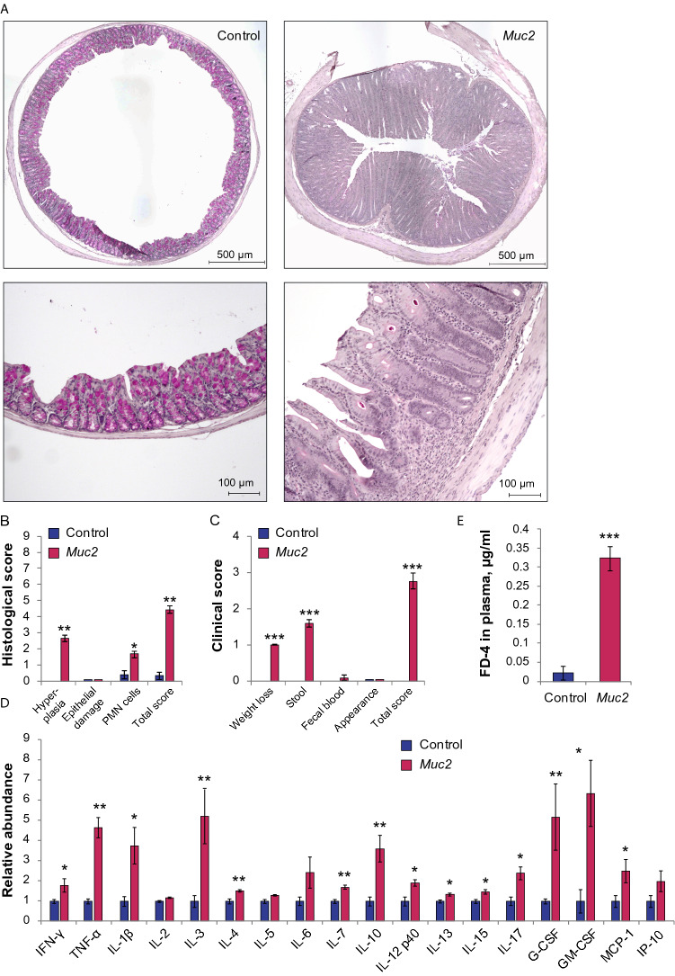
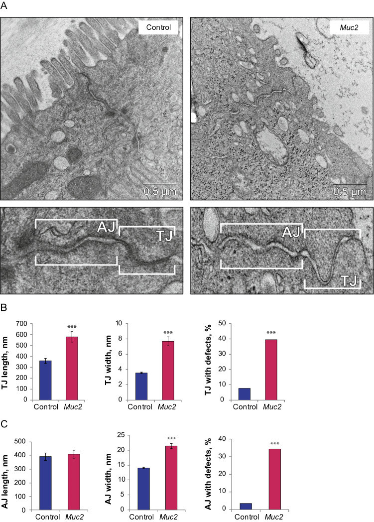
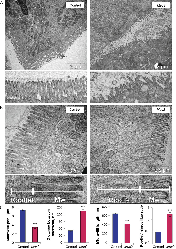
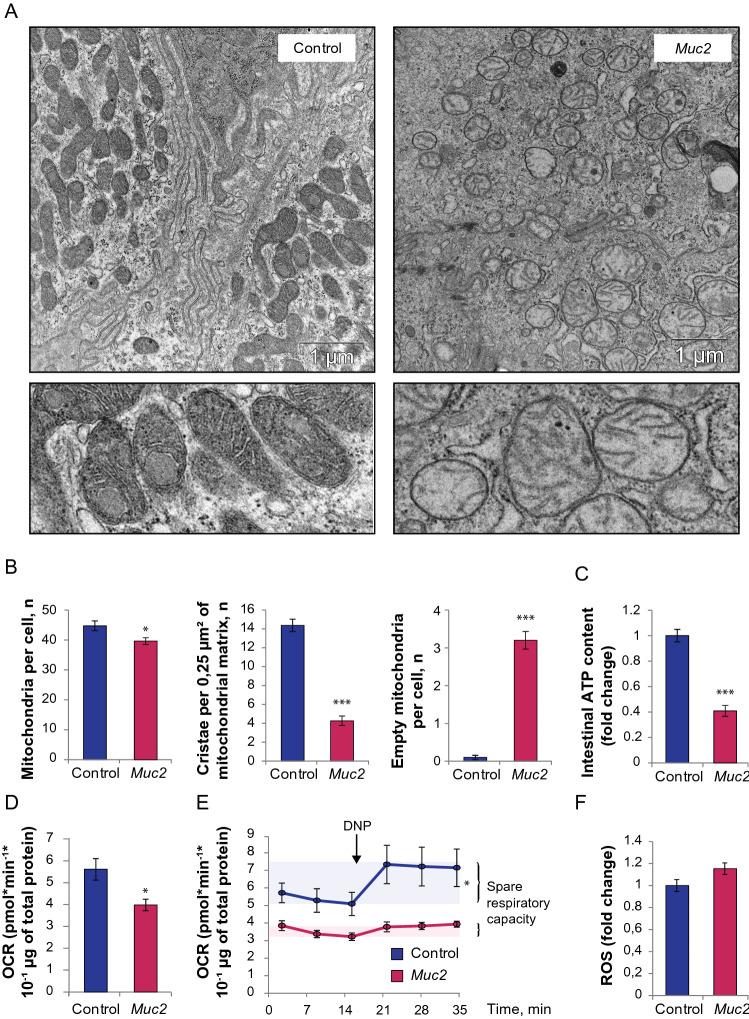
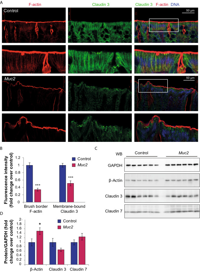
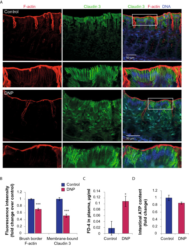
The disruption of the protective intestinal barrier-the 'leaky gut'-is a common complication of the inflammatory bowel disease. There is limited data on the mechanisms of the intestinal barrier disruption upon low-grade inflammation characteristic of patients with inflammatory bowel disease in clinical remission. Thus, animal models that recapitulate the complexity of chronic intestinal inflammation in vivo are of particular interest. In this study, we used Mucin-2 (Muc2) knockout mice predisposed to colitis to study intestinal barrier upon chronic inflammation. We used 4-kDa FITC-Dextran assay and transmission electron microscopy to demonstrate the increased intestinal permeability and morphological defects in intercellular junctions in Muc2 knockout mice. Confocal microscopy revealed the disruption of the apical F-actin cytoskeleton and delocalization of tight junction protein Claudin-3 from the membrane. We further demonstrate mitochondrial damage, impaired oxygen consumption and the reduction of the intestinal ATP content in Muc2 knockout mice. Finally, we show that chemically induced mitochondrial uncoupling in the wild type mice mimics the intestinal barrier disruption in vivo and causes partial loss of F-actin and membrane localization of Claudin-3. We propose that mitochondrial damage and metabolic shifts during chronic inflammation contribute to the leaky gut syndrome in Muc2 knockout animal model of colitis.


Conflict of interest statement

The authors declare no competing interests.

Figures

Figure 1
Figure 1
Muc2 knockout as a model of chronic intestinal inflammation and leaky gut. (A) PAS-stained colonic sections of C57Bl/6 and Muc2−/− mice. (B) Histological scoring of the inflammatory response. *p < 0.05, **p < 0.01, vs C57Bl/6, Mann–Whitney u-test. (C) Clinical scoring of the inflammatory response. *p < 0.05, **p < 0.01, vs C57Bl/6, Mann–Whitney u-test. (D) Cytokine levels in the descending colon. *p < 0.05, ** = p < 0.01, ***p < 0.001, vs C57Bl/6, Mann–Whitney u-test. (E) Intestinal permeability. ***p < 0.001, vs C57Bl/6, Student’s t-test.
Figure 2
Figure 2
Increased intestinal permeability results from lack of structural integrity in TJs. (A) TEM of the descending colon epithelium of C57Bl/6 and Muc2−/− mice. (B) TJ length (***p < 0.001, Mann–Whitney u test), width (***p < 0.001, Mann–Whitney u test) and the percentage of the defective TJs (***p < 0.001, χ2 test). (C) AJ length, width (***p < 0.001, Student’s t-test) and the percentage of the defective AJs (***p < 0.001, χ2 test).
Figure 3
Figure 3
Defective microvilli upon Muc2 knockout. (A) TEM of the microvilli in the descending colon epithelium of C57Bl/6 and Muc2−/− mice. (B) The detailed view of the microvilli structure. (C) Morphometric analysis: microvilli number per 1 µm of cell surface (***p < 0.001, Mann–Whitney u test), distance between microvilli (***p < 0.001, Mann–Whitney u test), microvillus length (***p < 0.001, Student’s t-test), rootlet/microvillus length ratio (***p < 0.001, Student’s t-test). Mw microvillus.
Figure 4
Figure 4
Mitochondrial damage and the reduction of ATP content and OCR in the descending colon in Muc2 knockout. (A) TEM of the mitochondria in the descending colon epithelium of C57Bl/6 and Muc2−/− mice. (B) Morphometric analysis: number of mitochondria per cell (*p < 0.05, vs C57Bl/6, Student’s t-test), number of cristae per 0.25 μm2 of mitochondrial matrix, ***p < 0.001, vs C57Bl/6, Student’s t-test), number of empty mitochondria per cell (***p < 0.001, vs C57Bl/6, Mann–Whitney u-test). (C) ATP level in the descending colon. ***p < 0.001, vs C57Bl/6, Student’s t-test. (D) Baseline OCR in the mitochondria of the isolated colonic crypts (*p < 0.05, vs C57Bl/6, Mann–Whitney u-test). (E) Spare respiratory capacity determined upon DNP treatment of the isolated colonic crypts (*p < 0.05 for C57Bl/6, Friedman test). (F) ROS formation in the isolated colonic crypts.
Figure 5
Figure 5
Lack of F-actin polymerization at the epithelial cell surface and Claudin-3 delocalization upon Muc2 knockout. (A) Claudin-3 and F-actin double immunostaining in the descending colon of C57Bl/6 and Muc2−/− mice. Maximum intensity projections through 40 µm of tissue are shown for each image. (B) Fluorescence intensity quantification of Claudin 3 along the cell membrane and F-actin within the brush border (***p < 0.001, vs C57Bl/6, Student’s t-test). (C) Western blot analysis of the total protein in colonic samples. (D) Quantification of the Western blot data (normalized to GAPDH protein level, fold change), *p < 0.05, vs C57Bl/6, Mann–Whitney u-test.
Figure 6
Figure 6
Chemically induced mitochondrial uncoupling in the C57Bl/6 mice induced the intestinal barrier disruption in vivo and decreased F-actin polymerization. (A) Claudin-3 and F-actin double immunostaining in the descending colon of C57Bl/6 mice, and in C57Bl/6 mice treated with 0.8 g/l DNP. Maximum intensity projections through 40 µm of tissue are shown for each image. (B) Fluorescence intensity quantification of Claudin 3 along the cell membrane and F-actin within the brush border (***p < 0.001, vs C57Bl/6, Student’s t-test). (C) Intestinal permeability (*p < 0.05, vs C57Bl/6, Student’s t-test). (D) ATP levels in the descending colon.

References

    1. Ivanov AI, Parkos CA, Nusrat A. Cytoskeletal regulation of epithelial barrier function during inflammation. Am. J. Pathol. 2010;177:512–524. doi: 10.2353/ajpath.2010.100168. - DOI - PMC - PubMed
    1. Zeissig S, et al. Changes in expression and distribution of claudin 2, 5 and 8 lead to discontinuous tight junctions and barrier dysfunction in active Crohn’s disease. Gut. 2007;56:61–72. doi: 10.1136/gut.2006.094375. - DOI - PMC - PubMed
    1. Schmitz H, et al. Altered tight junction structure contributes to the impaired epithelial barrier function in ulcerative colitis. Gastroenterology. 1999;116:301–309. doi: 10.1016/S0016-5085(99)70126-5. - DOI - PubMed
    1. Bruewer M, Samarin S, Nusrat A. Inflammatory bowel disease and the apical junctional complex. Ann. N. Y. Acad. Sci. 2006;1072:242–252. doi: 10.1196/annals.1326.017. - DOI - PubMed
    1. Buckley A, Turner JR. Cell biology of tight junction barrier regulation and mucosal disease. Cold Spring Harb. Perspect. Biol. 2018;10:a029314. doi: 10.1101/cshperspect.a029314. - DOI - PMC - PubMed

Publication types

LinkOut - more resources