. 2020 Dec 3;3(1):730.
doi: 10.1038/s42003-020-01474-3.

Ubiquitination of phytoene synthase 1 precursor modulates carotenoid biosynthesis in tomato

Affiliations

Ubiquitination of phytoene synthase 1 precursor modulates carotenoid biosynthesis in tomato

Peiwen Wang et al. Commun Biol. .

Abstract

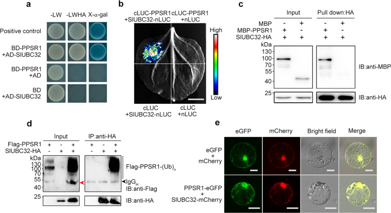
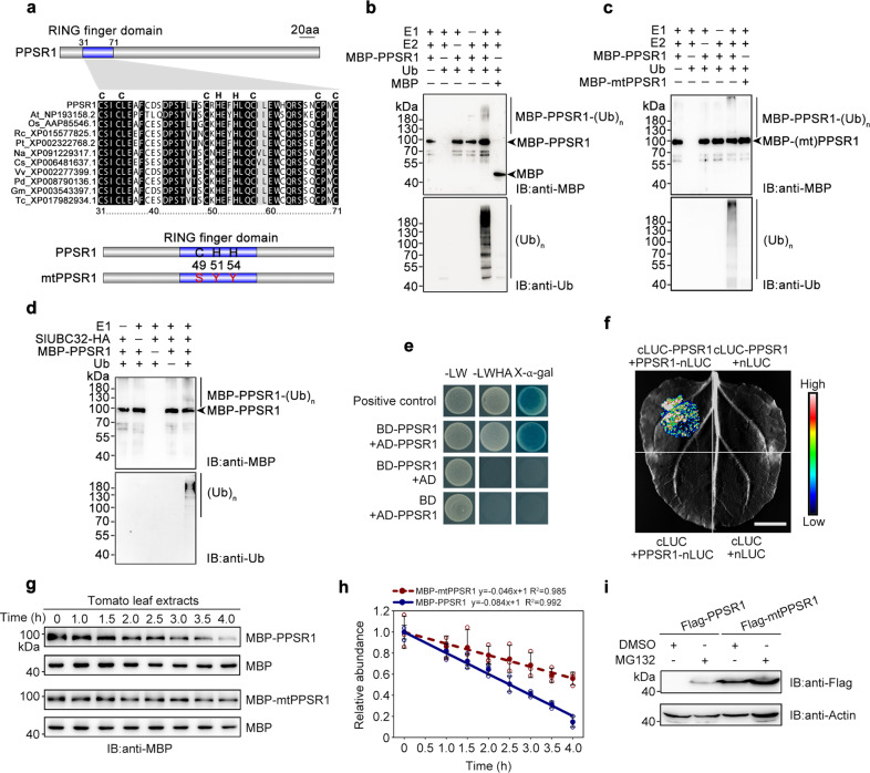
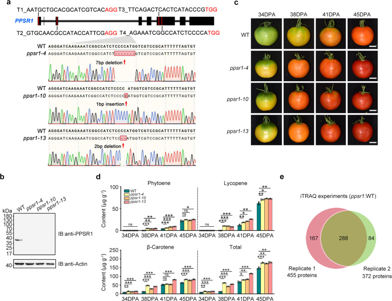
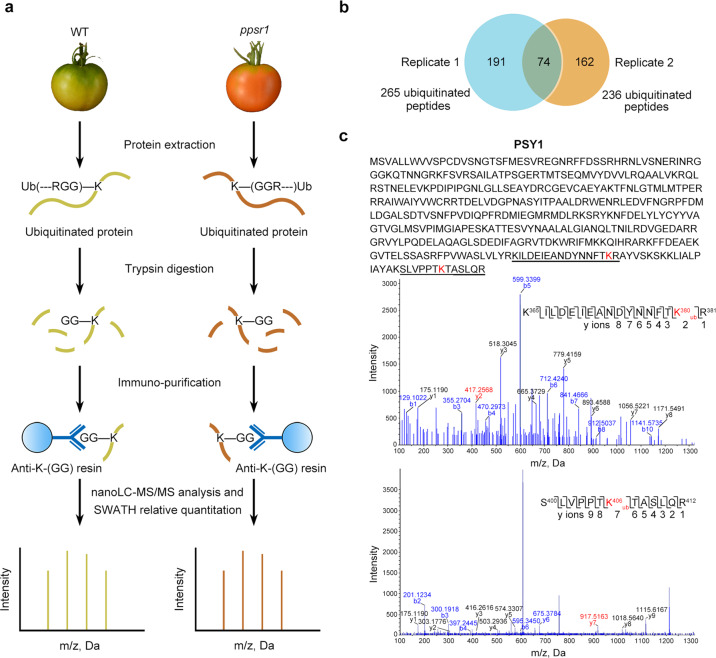
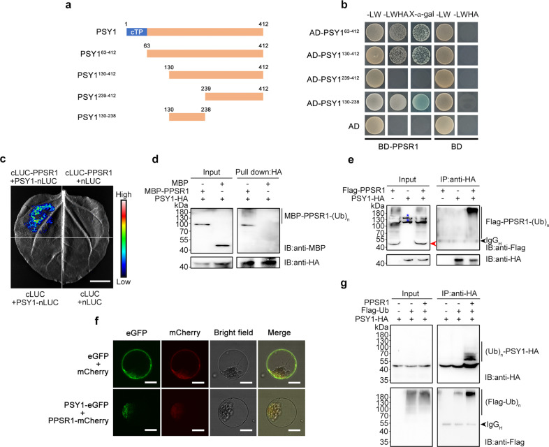
Carotenoids are natural pigments that are indispensable to plants and humans, whereas the regulation of carotenoid biosynthesis by post-translational modification remains elusive. Here, we show that a tomato E3 ubiquitin ligase, Plastid Protein Sensing RING E3 ligase 1 (PPSR1), is responsible for the regulation of carotenoid biosynthesis. PPSR1 exhibits self-ubiquitination activity and loss of PPSR1 function leads to an increase in carotenoids in tomato fruit. PPSR1 affects the abundance of 288 proteins, including phytoene synthase 1 (PSY1), the key rate-limiting enzyme in the carotenoid biosynthetic pathway. PSY1 contains two ubiquitinated lysine residues (Lys380 and Lys406) as revealed by the global analysis and characterization of protein ubiquitination. We provide evidence that PPSR1 interacts with PSY1 precursor protein and mediates its degradation via ubiquitination, thereby affecting the steady-state level of PSY1 protein. Our findings not only uncover a regulatory mechanism for controlling carotenoid biosynthesis, but also provide a strategy for developing carotenoid-enriched horticultural crops.


Conflict of interest statement

The authors declare no competing interests.

Figures

Fig. 1
Fig. 1. PPSR1 interacts with SlUBC32.
a Y2H assay revealing the interactions between PPSR1 and SlUBC32. The PPSR1 fused with the binding domain (BD) of GAL4 (BD-PPSR1) and the SlUBC32 fused with the activation domain (AD) of GAL4 (AD-SlUBC32) were co-expressed in yeast. The transformants were selected on SD/-Leu/-Trp (-LW) and SD/-Leu/-Trp/-His/-Ade (-LWHA) with or without X-α-gal. b LCI assay revealing the interactions between PPSR1 and SlUBC32. The PPSR1 fused with the C-terminus of LUC (cLUC-PPSR1) was co-expressed with the SlUBC32 fused with the N-terminus of LUC (SlUBC32-nLUC) in tobacco (Nicotiana benthamiana) leaves. Scale bar, 1 cm. c Pull-down assay revealing the interactions between PPSR1 and SlUBC32. The recombinant SlUBC32-HA, MBP-PPSR1, and MBP (negative control) were mixed as indicated, and incubated with anti-HA agarose. The eluted proteins were detected by immunoblot using anti-MBP and anti-HA antibodies, respectively. IB, immunoblot. d Co-IP assay revealing the interactions between PPSR1 and SlUBC32. The Flag-PPSR1 and SlUBC32-HA fusion proteins were co-expressed in N. benthamiana leaves. The total proteins were extracted from the infected leaves treated with MG132 and immunoprecipitated by anti-HA agarose. The eluted proteins were then detected by immunoblot using anti-Flag and anti-HA antibodies, respectively. The red arrowhead indicates the predicted Flag-PPSR1. The black arrowhead refers to heavy chain of antibody (IgGH). (Ub)n, polyubiquitin chain. e Subcellular colocalization of PPSR1 and SlUBC32. The Agrobacteria carrying 35S::PPSR1-eGFP and 35S::SlUBC32-mCherry constructs were transiently co-transformed into tobacco leaves. The tobacco protoplasts co-expressing eGFP and mCherry were used as negative control. Scale bars, 20 µm.
Fig. 2
Fig. 2. PPSR1 exhibits self-ubiquitination activity.
a Characterization of the RING finger domain in PPSR1. Sequence alignment of the RING finger domain reveals the conserved amino acids. The mutated form of PPSR1 (mtPPSR1) was generated by site-directed mutagenesis. C, cysteine; H, histidine; S, serine; Y, tyrosine. At, Arabidopsis thaliana; Os, Oryza sativa; Rc, Ricinus communis; Pt, Populus trichocarpa; Na, Nicotiana attenuata; Cs, Citrus sinensis; Vv, Vitis vinifera; Pd, Phoenix dactylifera; Gm, Glycine max; Tc, Theobroma cacao. b, c Ubiquitination assay of PPSR1 in vitro. An ubiquitination reaction was carried out in the presence (+) or absence (−) of E1, E2, ubiquitin (Ub), and MBP-PPSR1 (b) or MBP-mtPPSR1 (c). The reaction products were subjected to immunoblot using anti-MBP and anti-Ub antibodies, respectively. MBP protein was used as the negative control in (b). (Ub)n, polyubiquitin chain. IB, immunoblot. d PPSR1 ubiquitination assay using SlUBC32 instead of E2 used in (b). e Y2H assay revealing the self-interaction of PPSR1. The PPSR1 fused with the activation domain (AD) of GAL4 (AD-PPSR1) and the binding domain (BD) of GAL4 (BD-PPSR1) were co-expressed in yeast. The transformants were selected on SD/-Leu/-Trp (-LW) and SD/-Leu/-Trp/-His/-Ade (-LWHA) with or without X-α-gal. f LCI assay revealing the self-interaction of PPSR1. The PPSR1 fused with the C-terminus of LUC (cLUC-PPSR1) was co-expressed with the PPSR1 fused with the N-terminus of LUC (PPSR1-nLUC) in tobacco (Nicotiana benthamiana) leaves. Scale bar, 1 cm. g Cell-free degradation assay of PPSR1. The recombinant MBP-PPSR1 and MBP-mtPPSR1 proteins were purified and incubated in the extracts from tomato leaves, respectively. The protein levels were measured by immunoblot using an anti-MBP antibody at different time intervals. The MBP protein was used as the loading control. h Quantification of protein levels in (g) by ImageJ. Error bars represent the means ± standard deviation (SD) of three independent experiments. The circles indicate individual data points. i Stability analysis of PPSR1. The Flag-PPSR1 and Flag-mtPPSR1 fusion proteins were expressed in N. benthamiana leaves, respectively. The protein levels were determined by immunoblot using an anti-Flag antibody. The N. benthamiana actin was used as the loading control.
Fig. 3
Fig. 3. PPSR1 modulates carotenoid accumulation in tomato fruit during ripening.
a Genotyping of mutations mediated by CRISPR/Cas9 gene-editing system in ppsr1-4, ppsr1-10, and ppsr1-13 mutants. Schematic illustration shows the single guide RNAs (sgRNAs) containing different target sequences (T1, T2, T3, and T4) that were designed to specifically target the exons of PPSR1. Red letters represent the protospacer adjacent motif (PAM). Red arrows indicate the editing sites that were verified by sequencing. b Absence of PPSR1 protein in the ppsr1 mutants. Total proteins were extracted from fruit of wild-type (WT) and ppsr1 mutants at 38 days post-anthesis (DPA) and subjected to immunoblot using an anti-PPSR1 antibody. Equal loading was confirmed by an anti-actin antibody. IB, immunoblot. c Phenotype analysis of ppsr1 mutants. Fruit from WT and ppsr1 mutants at 34, 38, 41, and 45 DPA are shown. Scale bars, 2 cm. d Accumulation of carotenoids (phytoene, lycopene, and β-carotene) in fruit of WT and ppsr1 mutants during ripening. Error bars represent the means ± standard deviation (SD) of three independent experiments. The circles indicate individual data points. Asterisks indicate significant differences (*P < 0.05, **P < 0.01, ***P < 0.001; two-tailed Student’s t-test). e Venn diagram showing the overlap of proteins that exhibit differential expression in the ppsr1 mutant fruit compared to the WT in two independent biological replicates of quantitative proteome analysis. Proteins isolated from WT and ppsr1 mutant fruit at 38 DPA were subjected to iTRAQ (isobaric tags for relative and absolute quantification) labeling coupled with NanoLC–MS/MS.
Fig. 4
Fig. 4. Identification of PSY1 as the candidate substrate of PPSR1.
a A workflow diagram showing the identification and quantification of ubiquitinated peptides that exhibit significant differences in abundance in the ppsr1 mutant fruit compared to the wild type (WT). Proteins isolated from WT and ppsr1 mutant fruit at 38 DPA were digested with trypsin, followed by immunoprecipitation with an anti-K-(GG) antibody. The recovered ubiquitinated peptides were submitted to SWATH-MS (Sequential Window Acquisition of all Theoretical Mass Spectra) quantitative proteomic analysis. b Overlap of the ubiquitinated peptides identified in two independent biological replicates. c Identification of ubiquitination sites in PSY1 by NanoLC–MS/MS. Sequences of the identified ubiquitinated peptides are underlined in the PSY1 protein sequence. The mass spectra of two ubiquitinated peptides are displayed. The y-ions and the corresponding peptide sequence are presented, with ubiquitinated lysine (K) residue marked in red.
Fig. 5
Fig. 5. PPSR1 binds to PSY1 and mediates its ubiquitination.
a, b Y2H assay revealing the region of PSY1 that interacts with PPSR1. a Schematic illustration for full-length PSY1 protein and the truncated forms used in Y2H analysis. Numbers indicate the positions of the first and last amino acid in the sequences. cTP, chloroplast transit peptide. b The PPSR1 fused with the binding domain (BD) of GAL4 (BD-PPSR1) and the truncated PSY1 fused with the activation domain (AD) of GAL4 were co-expressed in yeast. The transformants were selected on SD/-Leu/-Trp (-LW) and SD/-Leu/-Trp/-His/-Ade (-LWHA) with or without X-α-gal. c LCI assay revealing the interactions between PSY1 and PPSR1. The PPSR1 fused with the C-terminus of LUC (cLUC-PPSR1) was co-expressed with the PSY1 fused with the N-terminus of LUC (PSY1-nLUC) in tobacco (Nicotiana benthamiana) leaves. Scale bar, 1 cm. d Semi-in vivo pull-down assay revealing the interactions between PPSR1 and PSY1. The recombinant MBP-PPSR1 and MBP (negative control) were mixed with PSY1-HA expressed in tobacco leaves, and incubated with anti-HA agarose. The eluted proteins were detected by immunoblot using anti-MBP and anti-HA antibodies, respectively. IB, immunoblot. e Co-IP assay revealing the interactions between PSY1 and PPSR1. The Flag-PPSR1 and PSY1-HA fusion proteins were co-expressed in tobacco leaves. f Subcellular colocalization of PSY1 and PPSR1. The PSY1-eGFP and PPSR1-mCherry fusion proteins were transiently co-expressed into tobacco leaves. The tobacco leaves expressing eGFP or mCherry were used as the negative control. Scale bars, 20 µm. g Ubiquitination assay of PSY1. The Agrobacteria carrying 35S::PSY1-HA, 35S::Flag-ubiquitin (Ub), and 35S::PPSR1 constructs were infiltrated into the tobacco leaves. For (e) and (g), the total proteins were extracted from the infected leaves treated with MG132 and incubated with anti-HA agarose to enrich PSY1-HA. The eluted proteins were subjected to immunoblot using anti-Flag and anti-HA antibodies, respectively. The red arrowhead indicates the predicted Flag-PPSR1. The black arrowhead refers to heavy chain of antibody (IgGH). Blue asterisks refer to nonspecific bands. IB, immunoblot; (Ub)n, polyubiquitin chain.
Fig. 6
Fig. 6. PPSR1 modulates PSY1 protein level via ubiquitination.
a Stability assay of PSY1. The PSY1-HA fusion protein was co-expressed with Flag-PPSR1 in tobacco (Nicotiana benthamiana) leaves. The total proteins were extracted and submitted to immunoblot using anti-HA and anti-Flag antibodies, respectively. The N. benthamiana actin was used as the loading control. IB, immunoblot. b Degradation rate assays of PSY1 under the action of PPSR1. The PSY1-HA and Flag-PPSR1 fusion proteins were co-expressed in tobacco leaves. The leaves were treated with translation inhibitor cycloheximide (CHX), and the total proteins were extracted for immunoblotting with anti-HA antibody at an indicated time point after treatment. c Quantification of protein levels in (b) by ImageJ. d Diagram showing PSY1 with ubiquitination site mutations. K, lysine; R, arginine. e Degradation rate assays of PSY1 and its mutated forms. The PSY1 and its mutated forms expressed in tobacco leaves were treated with CHX and submitted to immunoblot as described in (b). f Quantification of protein levels in (e) by ImageJ. g Expression of PSY1 protein in fruit of wild-type (WT) and ppsr1 mutants. Total protein was extracted and submitted to immunoblot using anti-PSY1 antibody (upper panel). Equal loading was confirmed by an anti-actin antibody. The protein levels were quantified by ImageJ (lower panel). h Gene expression of PSY1 in fruit of WT and ppsr1 mutants. Total RNA was isolated and submitted to quantitative real-time PCR. The ACTIN gene was used as the internal control. For (c), (f), (g), and (h), error bars represent the means ± standard deviation (SD) of three independent experiments. The circles indicate individual data points. Asterisks indicate significant differences (**P < 0.01, ***P < 0.001; two-tailed Student’s t-test). i The working model for PPSR1-mediated post-translational regulation of PSY1. PPSR1 directly interacts with SlUBC32 and mediates degradation of PSY1 precursor, which is nucleus-encoded and synthesized in the cytosol, via 26S proteasome. In the absence of PPSR1, more PSY1 precursor was transported into the plastid, leading to the accumulation of PSY1 protein. The increased PSY1 protein accelerates the biosynthesis of carotenoid in the plastid.

References

    1. Nisar N, Li L, Lu S, Khin NC, Pogson BJ. Carotenoid metabolism in plants. Mol. Plant. 2015;8:68–82. doi: 10.1016/j.molp.2014.12.007. - DOI - PubMed
    1. Yazdani M, et al. Ectopic expression of ORANGE promotes carotenoid accumulation and fruit development in tomato. Plant Biotechnol. J. 2019;17:33–49. doi: 10.1111/pbi.12945. - DOI - PMC - PubMed
    1. Cazzonelli CI, Pogson BJ. Source to sink: regulation of carotenoid biosynthesis in plants. Trends Plant Sci. 2010;15:266–274. doi: 10.1016/j.tplants.2010.02.003. - DOI - PubMed
    1. Llorente B, Martinez-Garcia JF, Stange C, Rodríguez-Concepción M. Illuminating colors: regulation of carotenoid biosynthesis and accumulation by light. Curr. Opin. Plant Biol. 2017;37:49–55. doi: 10.1016/j.pbi.2017.03.011. - DOI - PubMed
    1. Liu Y, et al. Manipulation of light signal transduction as a means of modifying fruit nutritional quality in tomato. Proc. Natl Acad. Sci. USA. 2004;101:9897–9902. doi: 10.1073/pnas.0400935101. - DOI - PMC - PubMed

Publication types

MeSH terms

Substances