. 2020 Jul-Sep;30(3):327-333.
doi: 10.4103/ijri.IJRI_207_20. Epub 2020 Oct 15.

Zuska's breast disease: Breast imaging findings and histopathologic overview

Affiliations

Zuska's breast disease: Breast imaging findings and histopathologic overview

Luis F Serrano et al. Indian J Radiol Imaging. 2020 Jul-Sep.

Abstract

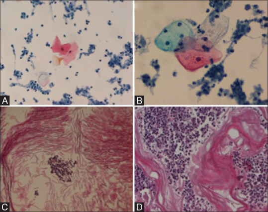
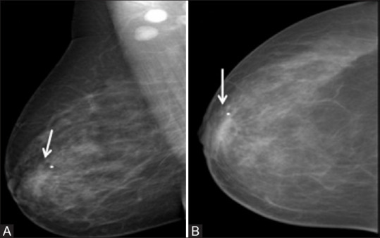
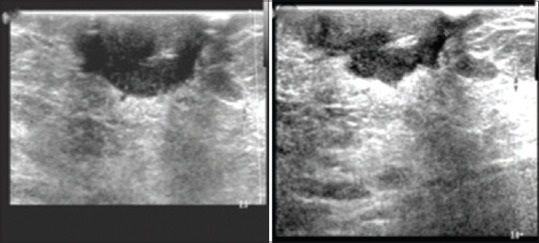
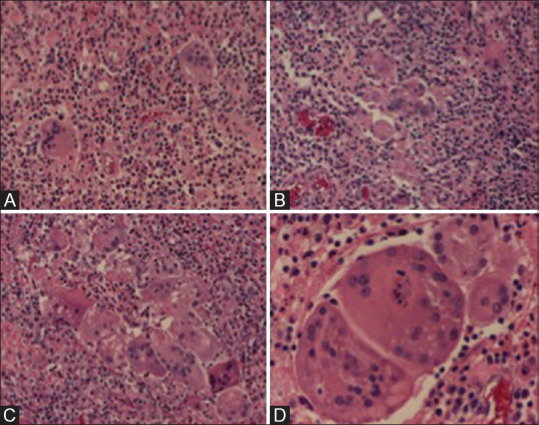
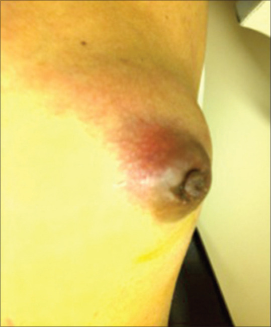
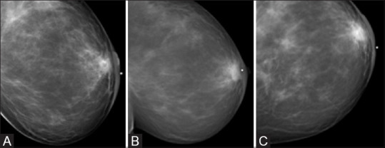
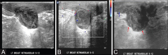
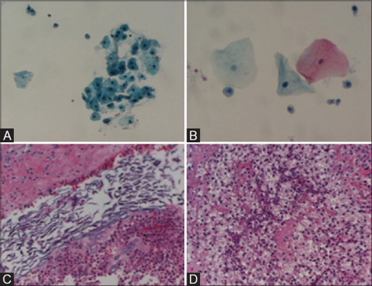
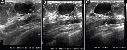
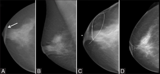
Zuska's disease describes the clinical condition of recurrent central or periareolar nonpuerperal abscesses associated with lactiferous fistulas. Pathogenesis involves the occlusion of an abnormal duct through an epithelial desquamation process that causes ductal dilatation, stasis of secretions, and periductal inflammation. Patients with Zuska's disease may develop chronic draining sinuses near the areola from lactiferous ducts fistula; therefore, the underlying abnormal duct system must be located and excised for proper treatment. Zuska's disease is often misdiagnosed and mistreated and is associated with significant morbidity, including the recurrence of abscess and cutaneous fistula formation. This case series aimed to help clinicians investigate and manage this disorder. The clinical and imaging findings, histopathologic correlation, and treatment of Zuska's disease are discussed.

Keywords: Abscess; Zuska's disease; breast diseases; lactiferous fistula.


Conflict of interest statement

There are no conflicts of interest.

Figures

Figure 1(<b>A-D</b>)
Figure 1(A-D)
Case 1. Contrast-enhanced CT images. (A and B) Axial plane. (C and D) Coronal plane. Arrows show a right retroareolar lobulated hyperdense mass measuring 3.2 cm × 3.2 cm
Figure 2(<b>A-C</b>)
Figure 2(A-C)
Case 1. Right breast mammogram. (A) Craniocaudal view (CC). (B) Mediolateral oblique view (MLO). (C) Lateral view. Retroareolar lobulated dense mass (White arrow) with skin thickening (Red arrow)
Figure 3
Figure 3
Case 1. Right breast ultrasound. Subareolar lobulated well-circumscribed mass with internal echoes
Figure 4(<b>A and B</b>)
Figure 4(A and B)
Case 1. PET scan. (A) Axial (B) Coronal. Right breast mass with increase uptake
Figure 5(<b>A-D</b>)
Figure 5(A-D)
Case 1. Histopathological examination. (A) FNA, filter preparation, Papanicolaou stain 200× magnification, mature squamous cells, and acute inflammatory exudate. (B) FNA, filter preparation, Papanicolaou stain, 400× magnification, mature squamous cells, and acute inflammatory exudate. (C) FNA, cell block preparation, H and E stain, 200× magnification, lamellated keratinous debris, and acute inflammatory exudate. (D) Core needle biopsy, H and E stain, 100× magnification, lamellated keratinous debris, and acute inflammatory exudate
Figure 6(<b>A-C</b>)
Figure 6(A-C)
Case 2. Abdomen CT performed for liver mass evaluation showing a right retroareolar breast mass. (A) Axial (B) Sagittal (C) Coronal
Figure 7(<b>A and B</b>)
Figure 7(A and B)
Case 2. Right breast mammogram. (A) MLO and (B) CC views, retroareolar asymmetric density and abnormal rounded dense lymph nodes in the ipsilateral axilla
Figure 8
Figure 8
Case 2. Right breast ultrasound. Multilobulated heterogeneous retroareolar mass with internal echoes
Figure 9(<b>A-D</b>)
Figure 9(A-D)
Case 2. Biopsy H and E stain. (A) 100X magnification. (B-C) 200 X magnification. (D) 400X magnification. Mixed inflammatory infiltrates and foreing-body-type multinucleated giant cells
Figure 10
Figure 10
Case 3. Large superficial mobile mass in the left areola
Figure 11(<b>A-C</b>)
Figure 11(A-C)
Case 3. Left breast mammogram. (A) CC, (B) MLO, and (C) Lateral views. Retroareolar asymmetry with areolar and skin thickening
Figure 12(<b>A-C</b>)
Figure 12(A-C)
Case 3. (A) Left breast ultrasound reveals a large hypoechoic irregular mass. (B) There is a mild increase in peripheral vascularity on color Doppler images. (C) Hypoechoic irregular mass (red arrows) beneath the skin with marked skin thickening (blue arrows)
Figure 13(<b>A-D</b>)
Figure 13(A-D)
Case 3. (A) FNA, filter preparation, Papanicolaou stain, 200× magnification, mature squamous cells, and scant neutrophils. (B) FNA, filter preparation, Papanicolaou stain, 400× magnification, mature squamous cells, and scant neutrophils. (C) Biopsy, H and E stain, 200× magnification, keratinous material and acute inflammatory infiltrate. (D) Biopsy, H and E stain, 200× magnification, and mixed inflammatory infiltrate with abundant foamy macrophages
Figure 14
Figure 14
Case 4. (A-C) Right breast ultrasound shows a well-circumscribed oval mass with dilated ducts (white arrows on A-C)
Figure 15(<b>A-D</b>)
Figure 15(A-D)
Case 4. Right breast mammogram. (A) CC and (B) MLO views. Subareolar skin thickening (arrow). The mass is obscured by dense glandular tissue. (C) Needle wire localization (D) Postoperative (mass removed, scar marker)

References

    1. Kasales CJ, Han B, Smith JS, Chetlen AL, Kaneda HJ, Shereef S. Nonpuerperal mastitis and subareolar abscess of the breast. Am J Roentgenol. 2014;202:W133–9. - PubMed
    1. Trop I, Dugas A, Davis J, El Khoury M, Boileau JF, Larouche N, et al. Breast abscesses: Evidence-based algorithms for diagnosis, management, and follow-up. Radiographics. 2011;31:1683–99. - PubMed
    1. Zuska JJ, Crile G, Ayres WW. Fistulas of lactifierous ducts. Am J Surg. 1951;81:312–7. - PubMed
    1. Livingston SF, Arlen M. Ductal fistula of the breast. Ann Surg. 1962;155:316–9. - PMC - PubMed
    1. Gollapalli V, Liao J, Dudakovic A, Sugg SL, Scott-Conner CE, Weigel RJ. Risk factors for development and recurrence of primary breast abscesses. J Am Coll Surg. 2010;211:41–8. - PubMed