Review
. 2020 Nov 15:2020:5021694.
doi: 10.1155/2020/5021694. eCollection 2020.

Oxidative Stress in Amyotrophic Lateral Sclerosis: Pathophysiology and Opportunities for Pharmacological Intervention

Affiliations
Review

Oxidative Stress in Amyotrophic Lateral Sclerosis: Pathophysiology and Opportunities for Pharmacological Intervention

Teresa Cunha-Oliveira et al. Oxid Med Cell Longev. .

Abstract

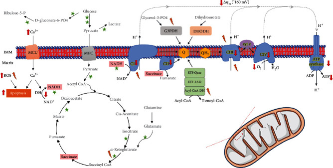
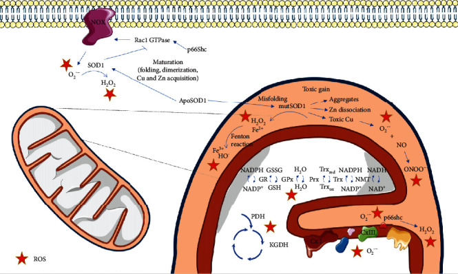
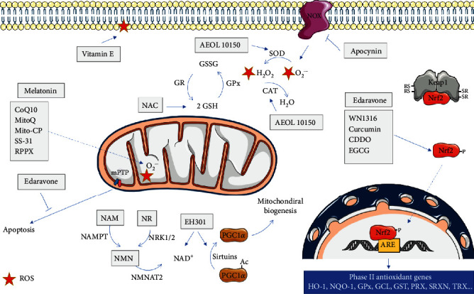
Amyotrophic lateral sclerosis (ALS), also known as Lou Gehrig's disease or Charcot disease, is a fatal neurodegenerative disease that affects motor neurons (MNs) and leads to death within 2-5 years of diagnosis, without any effective therapy available. Although the pathological mechanisms leading to ALS are still unknown, a wealth of evidence indicates that an excessive reactive oxygen species (ROS) production associated with an inefficient antioxidant defense represents an important pathological feature in ALS. Substantial evidence indicates that oxidative stress (OS) is implicated in the loss of MNs and in mitochondrial dysfunction, contributing decisively to neurodegeneration in ALS. Although the modulation of OS represents a promising approach to protect MNs from degeneration, the fact that several antioxidants with beneficial effects in animal models failed to show any therapeutic benefit in patients raises several questions that should be analyzed. Using specific queries for literature search on PubMed, we review here the role of OS-related mechanisms in ALS, including the involvement of altered mitochondrial function with repercussions in neurodegeneration. We also describe antioxidant compounds that have been mostly tested in preclinical and clinical trials of ALS, also describing their respective mechanisms of action. While the description of OS mechanism in the different mutations identified in ALS has as principal objective to clarify the contribution of OS in ALS, the description of positive and negative outcomes for each antioxidant is aimed at paving the way for novel opportunities for intervention. In conclusion, although antioxidant strategies represent a very promising approach to slow the progression of the disease, it is of utmost need to invest on the characterization of OS profiles representative of each subtype of patient, in order to develop personalized therapies, allowing to understand the characteristics of antioxidants that have beneficial effects on different subtypes of patients.


Conflict of interest statement

The authors declare no competing financial interest.

Figures

Figure 1
Figure 1
Mitochondrial dysfunction in sporadic forms of ALS. Mitochondrial bioenergetics is driven by the oxidation of different substrates and is stimulated by calcium. Flux of electrons through the electron transport chain creates a transmembrane proton gradient of about 160 mV in the resting state (negative inside), which fuels ATP synthesis in the mitochondrial matrix. Leak of electrons in some of the bioenergetic reactions generates reactive oxygen species (ROS) that are involved in important cellular signaling processes but that, when in excess, may also lead to cellular dysfunction and death. Fibroblasts from sALS patients showed markers of mitochondrial dysfunction, compared to control fibroblasts, including decreased activity of metabolic dehydrogenases, increased ROS levels, increased intracellular calcium levels, decreased expression of components of the oxidative phosphorylation system, decreased mitochondrial potential, oxygen consumption, and ATP levels [68]. Abbreviations: NAD: β-Nicotinamide adenine dinucleotide; NADH: β-Nicotinamide adenine dinucleotide 2′-phosphate reduced form; FAD: Flavin Adenine Dinucleotide; CI: Complex I; CII: Complex II; CIII: Complex III; CIV: Complex IV; Cyt c: Cytochrome c; ETF: electron transfer flavoprotein; ROS: reactive oxygen species; DH: dehydrogenase; MCU: mitochondrial calcium uniporter; MPC: mitochondrial pyruvate carrier; ΔΨm: mitochondrial transmembrane electric potential; ATP: adenosine triphosphate; ADP: adenosine diphosphate; IMM: inner mitochondrial membrane
Figure 2
Figure 2
Mitochondrial dysfunction associated with SOD1 mutations. Reactive oxygen species (ROS) may be formed in several cellular reactions and are controlled by a network of antioxidant enzymes that include superoxide dismutase 1 (SOD1), a Cu-Zn metalloprotein responsible for the conversion of O2•- into O2 and H2O2, which is mainly localized in the cytosol. SOD1 mutations are one of the most studied causes of ALS. Mutant SOD1 (mutSOD1) toxic gain involves its translocation to the mitochondrial intermembrane space, where it aggregates due to lower stability of mutSOD1 monomers/dimers.mutSOD1 may also cause elevated oxidative damage through the dissociation of zinc from the enzyme or exposure to toxic copper at the active site, promoting reverse O2•- production. O2•- reacts with nitric oxide generated by nitric oxide synthase, more rapidly than it does with SOD1, producing peroxynitrite, with consequent tyrosine nitration of cellular proteins. mutSOD1 may also act as a peroxidase by using H2O2 as a substrate, or the H2O2 produced in the dismutation reaction may originate HO through the Fenton reaction. mutSOD1 may also induce the activation of p66Shc, a protein involved in controlling mitochondrial redox homeostasis. Outside mitochondria, mutSOD1 associates more strongly with Rac1 compared to the wild type form of SOD1, being less sensitive to redox uncoupling, consequently leading to an increase in NADPH oxidase- (NOX-) derived O2•-. ApoSOD1: metal-deficient Cu,Zn-superoxide dismutase; NADP: β-Nicotinamide adenine dinucleotide 2′-phosphate; NADPH: β-Nicotinamide adenine dinucleotide 2′-phosphate reduced form; NAD: β-Nicotinamide adenine dinucleotide; NADH: β-Nicotinamide adenine dinucleotide 2′-phosphate reduced form; GSH: reduced glutathione; GSSG: oxidized glutathione; Trxred: reduced Thioredoxin; Trxox: oxidized Thioredoxin; Trx: Thioredoxin, NMT: N-myristoyltransferase; Prx: peroxiredoxin; GPx: glutathione peroxidase; GR: glutathione reductase; PDH: pyruvate dehydrogenase; KGDH: alpha-ketoglutarate dehydrogenase; CxI: complex I; CxIII: complex III.
Figure 3
Figure 3
Mitochondrial effects of different antioxidant agents in ALS. The scheme represents the main molecular targets of antioxidants used in ALS, as discussed in the main text. HO-1: heme oxygenase 1; NQO-1: NADPH quinine oxidoreductase 1; GPx: glutathione peroxidase; GCL: γ-glutamylcysteine synthetase; GST: glutathione S-transferase; PRX: peroxiredoxin; SRXN: sulfiredoxin; TRX: Thioredoxin; GR: glutathione reductase; CAT: catalase; SOD: superoxide dismutase; NAMPT: nicotinamide phosphoribosyltransferase; NMNAT2: nicotinamide/nicotinic acid mononucleotide adenylyltransferase 2; NRK1/2: nicotinamide riboside kinase 1/2; CoQ10: coenzyme Q10; RPPX: dexpramipexole; NAC:N-acetyl cysteine; CDDO: 2-cyano-3,12-dioxooleana-1,9,-dien-28-oic acid; EGCG: epigallocatechin gallate; ROS: reactive oxygen species.

Similar articles

Cited by

References

    1. Tafuri F., Ronchi D., Magri F., Comi G. P., Corti S. SOD1 misplacing and mitochondrial dysfunction in amyotrophic lateral sclerosis pathogenesis. Frontiers in Cellular Neuroscience. 2015;9:p. 336. doi: 10.3389/fncel.2015.00336. - DOI - PMC - PubMed
    1. Zarei S., Carr K., Reiley L., et al. A comprehensive review of amyotrophic lateral sclerosis. Surgical Neurology International. 2015;6(1):p. 171. doi: 10.4103/2152-7806.169561. - DOI - PMC - PubMed
    1. Smith E. F., Shaw P. J., De Vos K. J. The role of mitochondria in amyotrophic lateral sclerosis. Neuroscience Letters. 2019;710, article 132933 doi: 10.1016/j.neulet.2017.06.052. - DOI - PubMed
    1. Tanaka M., Sakata T., Palumbo J., Akimoto M. A 24-week, phase III, double-blind, parallel-group study of edaravone (MCI-186) for treatment of amyotrophic lateral sclerosis (ALS) (P3.189) Neurology. 2016;86
    1. The Writing Group. Safety and efficacy of edaravone in well defined patients with amyotrophic lateral sclerosis: a randomised, double-blind, placebo-controlled trial. The Lancet Neurology. 2017;16(7):505–512. doi: 10.1016/S1474-4422(17)30115-1. - DOI - PubMed

MeSH terms