Review
. 2020 Dec;12(4):417-429.
doi: 10.4055/cios20060. Epub 2020 Nov 18.

Management of Osteogenesis Imperfecta: A Multidisciplinary Comprehensive Approach

Affiliations
Review

Management of Osteogenesis Imperfecta: A Multidisciplinary Comprehensive Approach

Tae-Joon Cho et al. Clin Orthop Surg. 2020 Dec.

Abstract

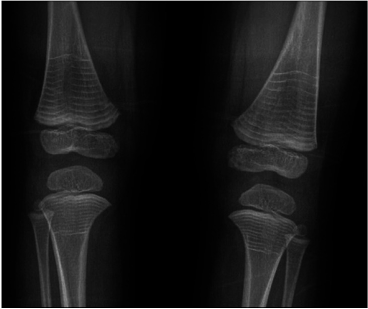
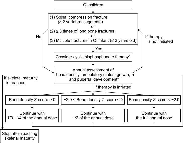
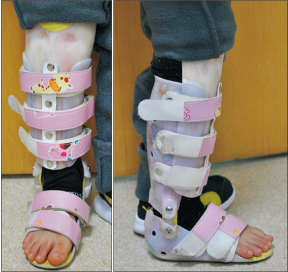
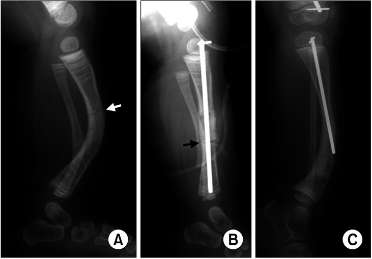
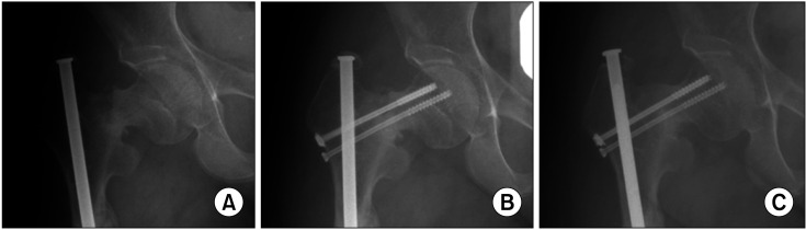
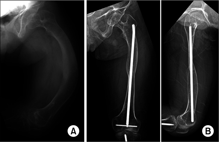
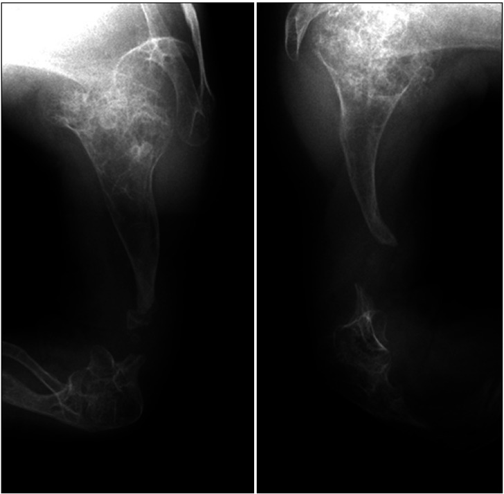
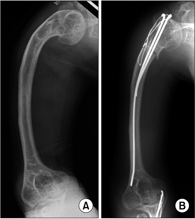
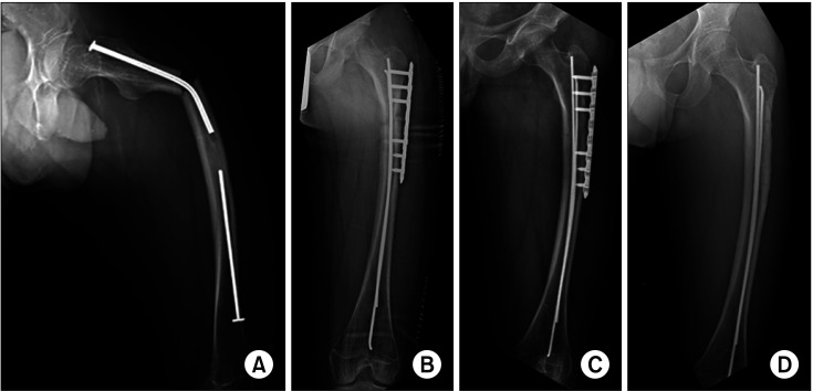
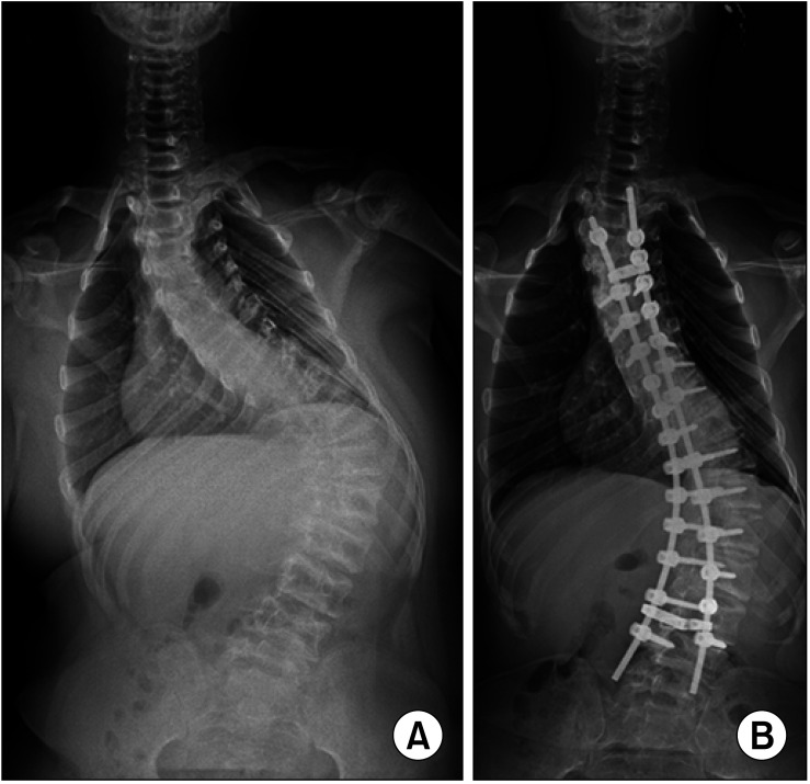
Osteogenesis imperfecta (OI) is characterized by recurring fractures and limb and spine deformities. With the advent of medical therapeutics and the discovery of causative genes, as well as the introduction of a newly devised intramedullary rod, the general condition and ambulatory function of patients diagnosed with OI have been improved over the past decades. This review covers recent developments in research and management of OI.

Keywords: Bisphosphonate; Gene; Intramedullary nailing; Osteogenesis imperfecta; Scoliosis.


Conflict of interest statement

CONFLICT OF INTEREST: No potential conflict of interest relevant to this article was reported.

Figures

Fig. 1
Fig. 1. Zebra lines at the metaphyses of long bones are formed by cyclic administration of pamidronate. Osteosclerotic lines parallel to the physis represent the primary spongiosa produced during each cycle of pamidronate infusion.
Fig. 2
Fig. 2. Protocol outlining cyclic bisphosphonate therapy for osteogenesis imperfecta (OI) patients. *If cyclic bisphosphonate therapy is decided, starting with the full annual dose is recommended. Pamidronate: 9 mg/kg/yr, 4–9 divided doses (max 60 mg/dose). Zoledronate: 0.1 mg/kg/yr, 2 divided doses (max 5 mg/yr). If bone density Z-score is > 2.0, running or sprinting is possible, normal height of vertebrae is preserved, or 0–2 times of long bone fractures occur per year, initiation of cyclic bisphosphonate therapy can be delayed with annual monitoring.
Fig. 3
Fig. 3. Patellar tendon-bearing total contact brace with hinged ankle.
Fig. 4
Fig. 4. (A) A 2.5-year-old girl with type III osteogenesis imperfecta sustained a linear fracture (white arrow) at the anteriorly angulated left tibia. (B) Additional osteotomy (black arrow) was performed to straighten the tibia, which was fixed with a non-elongating intramedullary rod because the tibia was too small to fit a telescopic rod. (C) Two years later, the tibia outgrew the rod and angulated at its distal tip.
Fig. 5
Fig. 5. (A) A 22-year-old woman with type IV OI showed delayed malunion of basicervical stress fracture. (B) Threaded screws were inserted and passed anterior to the preexisting telescoping rod. (C) Bony union was confirmed radiographically at the 1-year follow-up.
Fig. 6
Fig. 6. (A) A 19-year-old woman with type III OI manifested approximately 90° retroversion of the femur. The proximal femur appeared to be an anteroposterior (AP) projection, but the distal femur to be a lateral projection. (B) After extension and rotational osteotomy, the femur showed normal configuration on both AP and lateral projections.
Fig. 7
Fig. 7. Pseudarthrosis of the distal humerus in a 19-year-old man, who grew up in an orphanage. The elbow joint was stiff and all the motion occurred at the pseudarthrosis site, so the lesion was skillfully neglected.
Fig. 8
Fig. 8. Proximal humerus varus deformity in a 30-year-old woman with type III osteogenesis imperfecta (A) was corrected by closing wedge osteotomy fixed with tension band wiring (B). Two long flexible intramedullary rods were used to avoid a stress-riser effect.
Fig. 9
Fig. 9. Two cases of peri-implant fractures at the tip of plate screw: a 30-year-old man (A) and a 14-year-old boy (B).
Fig. 10
Fig. 10. (A) A 4-year-old boy was referred after his femoral shaft fracture was fixed with flexible intramedullary rods. (B) One rod was removed as it migrated distally. The remaining rod cut through the anterolateral cortex in both proximal and distal areas. The protruding tip irritated the overlying muscles, and eventually a fracture occurred at the distal cut-through point.
Fig. 11
Fig. 11. (A) Implantation of a Sheffield rod into the tibia. The T-piece of an obturator was introduced into the distal tibial epiphysis via ankle arthrotomy and through the articular cartilage. (B) Repeat arthrotomy of the knee joint showed the scar at the distal femoral articular cartilage following implantation of the obturator of Sheffield rod.
Fig. 12
Fig. 12. Schematic drawing of the surgical procedure for insertion of an interlocking telescopic rod in the tibia. The obturator is inserted within the sleeve in an antegrade manner and is transfixed at the distal epiphysis. Adapted from Cho et al. with permission of Wolters Kluwer Health.
Fig. 13
Fig. 13. (A) A 5-year-old boy with type IV osteogenesis imperfecta sustained a tibial shaft fracture and was treated with open reduction-additional osteotomy-interlocking telescopic intramedullary rod fixation. (B) Four years later, the female rod was replaced with a longer dual interlocking telescopic rod. (C) Until age 13 years, the rod telescoped successfully and continued to stabilize the tibia.
Fig. 14
Fig. 14. Telescopic rod designs. (From left) Dual Rush pins; Sheffield rod; Fassier-Duval rod; and interlocking telescopic rod of the authors' design.
Fig. 15
Fig. 15. (A) A 2-year-old girl with type IV osteogenesis imperfecta underwent Sofield operation on the right femur, which was fixed with a Sheffield rod. (B) The rod telescoped successfully for 3 years. (C) At age 7 years, the rod was bent at the proximal femur and failed to telescope. The T-piece of the obturator was located at the physis. (D) At the age of 8 years, the rod was further bent, and the T-piece of the obturator was located at the distal metaphysis.
Fig. 16
Fig. 16. (A) A 24-year-old woman with type IV osteogenesis imperfecta was referred due to nonunion of a distraction gap during femoral lengthening for height. (B) The distal femoral condyle collapsed, resulting in total stiff knee and severe disability in the patient.
Fig. 17
Fig. 17. (A) A 16-year-old boy with type I osteogenesis imperfecta sustained a subtrochanteric femur fracture. (B) Open reduction and internal fixation with 2 small flexible intramedullary (IM) rods and a locking plate with unicortical screws. (C) Bony union was achieved in 3 years, and the plate and screws were removed. (D) The IM rods were left behind by the age of 21 years.
Fig. 18
Fig. 18. (A) A 13-year-old boy with type V osteogenesis imperfecta underwent posterior spinal fusion with modern pedicle screw-based instrument for treatment of progressive scoliosis. (B) Five years later, a postoperative radiograph showed adequate spinal balance without further progression of deformity.

Similar articles

Cited by

References

    1. Cho TJ, Lee KE, Lee SK, et al. A single recurrent mutation in the 5′-UTR of IFITM5 causes osteogenesis imperfecta type V. Am J Hum Genet. 2012;91(2):343–348. - PMC - PubMed
    1. Semler O, Garbes L, Keupp K, et al. A mutation in the 5′-UTR of IFITM5 creates an in-frame start codon and causes autosomal-dominant osteogenesis imperfecta type V with hyperplastic callus. Am J Hum Genet. 2012;91(2):349–357. - PMC - PubMed
    1. Mortier GR, Cohn DH, Cormier-Daire V, et al. Nosology and classification of genetic skeletal disorders: 2019 revision. Am J Med Genet A. 2019;179(12):2393–2419. - PubMed
    1. Keupp K, Beleggia F, Kayserili H, et al. Mutations in WNT1 cause different forms of bone fragility. Am J Hum Genet. 2013;92(4):565–574. - PMC - PubMed
    1. Trejo P, Palomo T, Montpetit K, et al. Long-term follow-up in osteogenesis imperfecta type VI. Osteoporos Int. 2017;28(10):2975–2983. - PubMed

MeSH terms