Review
. 2021 Jan;13(2):99-116.
doi: 10.4155/fmc-2020-0288. Epub 2020 Dec 4.

Methods for the discovery of small molecules to monitor and perturb the activity of the human proteasome

Affiliations
Review

Methods for the discovery of small molecules to monitor and perturb the activity of the human proteasome

Marianne E Maresh et al. Future Med Chem. 2021 Jan.

Abstract

Regulating protein production and degradation is critical to maintaining cellular homeostasis. The proteasome is a key player in keeping proteins at the proper levels. However, proteasome activity can be altered in certain disease states, such as blood cancers and neurodegenerative diseases. Cancers often exhibit enhanced proteasomal activity, as protein synthesis is increased in these cells compared with normal cells. Conversely, neurodegenerative diseases are characterized by protein accumulation, leading to reduced proteasome activity. As a result, the proteasome has emerged as a target for therapeutic intervention. The potential of the proteasome as a therapeutic target has come from studies involving chemical stimulators and inhibitors, and the development of a suite of assays and probes that can be used to monitor proteasome activity with purified enzyme and in live cells.

Keywords: degradation; proteasome; screening.


Conflict of interest statement

Financial & competing interests disclosure

This work was supported through the Purdue University Center for Cancer Research NIH grant (P30 CA023168), the American Cancer Society Institutional Research Grant (IRG-14-190-56) to the Purdue University Center for Cancer Research, and a grant from the NIH-NIGMS (R21GM131206). Prof. Trader is a scientific co-founder and stock holder of Booster Therapeutics GmbH. The other authors have no other relevant affiliations or financial involvement with any organization or entity with a financial interest in or financial conflict with the subject matter or materials discussed in the manuscript apart from those disclosed.

No writing assistance was utilized in the production of this manuscript.

Figures
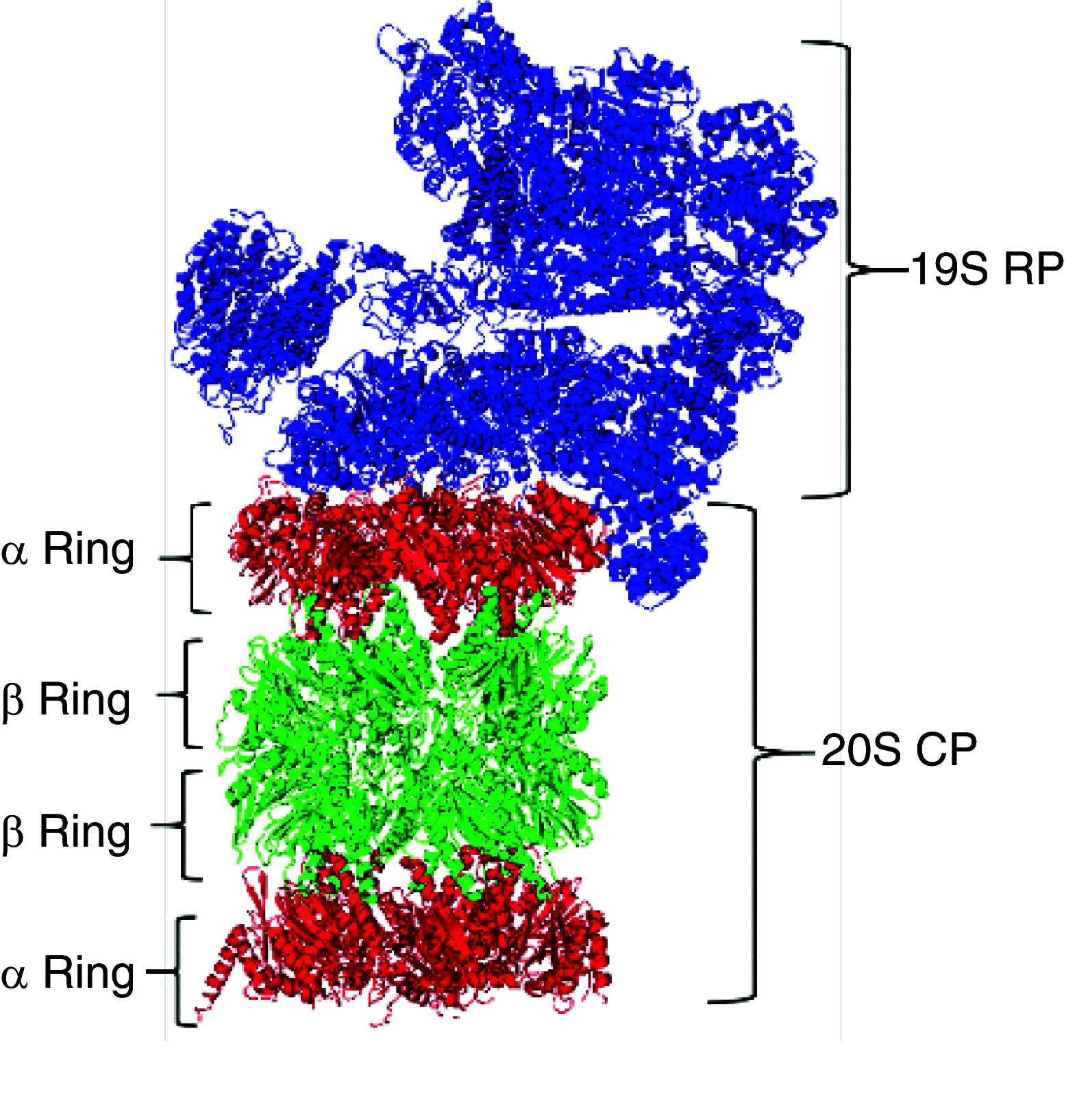
Figure 1.
Figure 1.. Structure of the 26S proteasome.
The 26S proteasome is composed of two parts, the 19S regulatory particle and the 20S catalytic core particle (sCP). The 19S particle is responsible for recognizing ubiquitinated substrates and shuttling them into the sCP. The sCP then hydrolyzes the protein into short peptide sequences that can be recycled by the cell. The sCP also exists in the uncapped form. In this state, it degrades small, disordered proteins.
Figure 2.
Figure 2.. Maintaining proteostasis is critical to cell viability.
The rate of protein synthesis must be matched by protein degradation to maintain proteostasis in the cell. If protein synthesis is too rapid, as is the case in several cancers, proteasomes are unable to degrade excess proteins efficiently, leading to protein accumulation and cell death. Similarly, if proteasome activity is suppressed (as is seen in some neurodegenerative diseases), proteins accumulate. To avoid death, cancer cells enhance proteasome activity by overexpressing genes encoding the proteasome subunits. Attention has also been turned to chemical stimulation of the proteasome as a mechanism to treat protein accumulation diseases (e.g., Parkinson disease) and pathologies related to aging.
Figure 3.
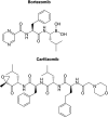
Figure 3.. Structures of bortezomib and carfilzomib.
Bortezomib and carfilzomib have both been approved by the US FDA for treatment of multiple myeloma. Bortezomib reversibly binds the β5 subunit via the boronic acid moiety. Carfilzomib irreversibly binds catalytic subunits via the epoxyketone moiety.
Figure 4.
Figure 4.. Broad spectrum proteasome inhibitor probes.
(A) AdaY(125I)Ahx3L3VS, a proteasome inhibitor that binds all catalytic subunits at concentrations greater than 1 μM. The vinyl sulfone moiety covalently binds catalytic threonine residues, and the radiolabeled iodine facilitates visualization of bound subunits after SDS-PAGE analysis. (B) MV151 was created using the same scaffold, but exchanging the radiolabel tag for BODIPY, a fluorescent tag. This new inhibitor has been demonstrated to retain the ability to covalently bind proteasome subunits while being easier to detect with fluorescent imaging after SDS-PAGE. It is also cell permeable. BODIPY: Boron-dipyrromethene; TMR: Tetramethylrhodamine.
Figure 5.
Figure 5.. Structures of subunit-specific probes.
Subunit-specific probes have enhanced the understanding of the proteasome and its activity in disease states such as cancer. NC-001 is specific to the β1/β1i subunits. LU-112 is specific to the β2/β2i subunit. NC-005 is the parent compound of NC-005-VS. Both of these inhibitors target the β5 subunit, but exchanging the epoxyketone of NC-005 for the vinyl sulfone shown in NC-005-VS provides more specificity to the β5 subunit. These probes have been conjugated to a variety of fluorophores and used to study the composition and activities of proteasomes in disease states and a multitude of cell types. Additionally, they have been used to monitor binding of small molecules to proteasome subunits.
Figure 6.
Figure 6.. Screening one-bead-one-compound libraries with magnetic beads.
The use of magnetic beads has enhanced the process of discovering new ligands to proteins of interest. Briefly, a protein incubates with beads of a one-bead-one-compound library, then a primary antibody against the protein is added and will only interact with beads bound to the protein. Magnetic beads coated with a secondary antibody are then added and bind only beads bound to the protein/primary antibody. A strong magnet is used to separate hits from non-hits and techniques such as LC/MS are used to identify the small molecule. LC/MS: Liquid chromatography mass spectrometry.
Figure 7.
Figure 7.. Structure of KDT-11.
KDT-11 was identified as part of a screen against a one-bead-one-compound library using magnetic beads. It binds Rpn-13 with a KD of ∼2 μM and is toxic to multiple myeloma cells.
Figure 8.
Figure 8.. Screening scheme for one-bead-one-compound libraries using fluorescently labeled Proteins.
Our new method to screen one-bead-one-compound libraries involves labeling a protein of interest with a near-infrared fluorophore. The protein is then incubated with beads from a one-bead-one-compound library that have been individually separated into the wells of a 96-well plate. Beads are then rinsed and the plate is imaged for fluorescence. The fluorescence intensities that result from the labeled protein binding the ligand on-bead can be quantified and provide a basis to rank hits.
Figure 9.
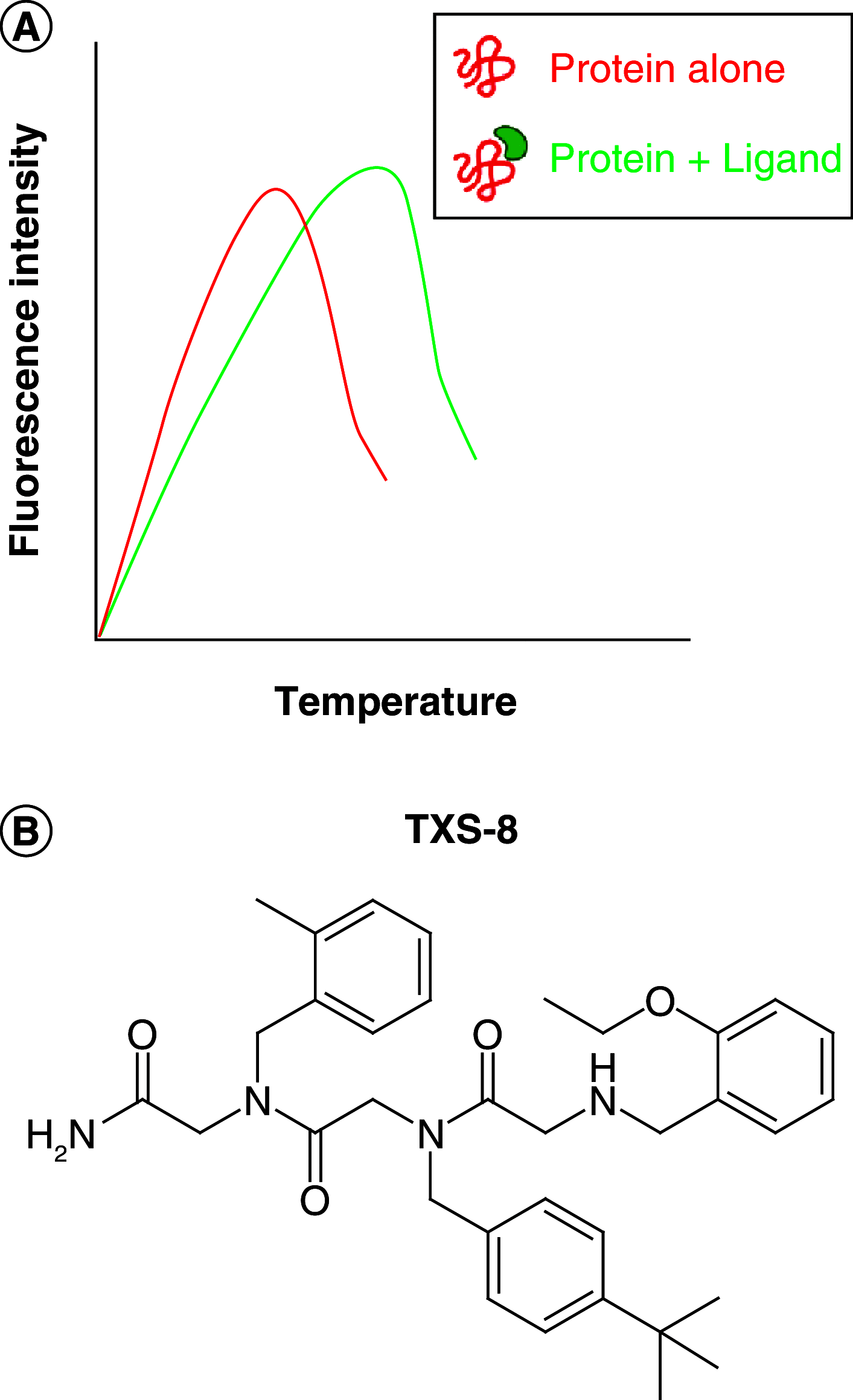
Figure 9.. Identification of TXS-8 through a thermal shift assay.
(A) Thermal shift assays rely on changes in the melting temperature of a protein in response to binding a small molecule. Fluorescent dyes that bind hydrophobic regions of proteins are used to monitor denaturation as the protein is heated. When a small molecule binds and stabilizes a protein, the melting temperature is increased and this shift can be quantified by measuring fluorescence intensity. (B) The structure of TXS-8, a peptoid binder of Rpn-6 identified from a thermal shift assay.
Figure 10.
Figure 10.. Structure of fluorogenic peptides used in high-throughput screening assays.
(A) Structure of the Suc-LLVY-AMC substrate. In red is highlighted the 7-amino-4-methylcoumarin scaffold. (B) Structure of the FRET peptide using EDANS (green) and DABCYL (magenta) as fluorescence pair. FRET: Fluorescence resonance energy transfer.
Figure 11.
Figure 11.. Newly developed probes to monitor proteasome activity.
(A) Structure of the TAS probes, highlighting the 20S isoform recognition sequence in green, where TAS1 contains a tyrosine residue (X: OH), TAS2 a 4-Cl phenylalanine residue (X: Cl) and TAS3 a 4-NO2 phenylalanine residue. (B) Structure of covalent proteasome activity probe that can react with all three active sites.

References

    1. Lecker SH, Goldberg AL, Mitch WE. Protein degradation by the ubiquitin–proteasome pathway in normal and disease states. J. Am. Soc. Nephrol. 17(7), 1807–1819 (2006). - PubMed
    1. Myung J, Kim KB, Crews CM. The ubiquitin-proteasome pathway and proteasome inhibitors. Med. Res. Rev. 21(4), 245–273 (2001). - PMC - PubMed
    1. Kloetzel PM. Antigen processing by the proteasome. Nat. Rev. Mol. Cell Biol. 2(3), 179–187 (2001). - PubMed
    1. Muli CS, Tian W, Trader DJ. Small-molecule inhibitors of the proteasome’s regulatory particle. ChemBioChem. 20(14), 1739–1753 (2019). - PMC - PubMed
    1. Bedford L, Paine S, Sheppard PW, Mayer RJ, Roelofs J. Assembly, structure, and function of the 26S proteasome. Trends Cell Biol. 20(7), 391–401 (2010). - PMC - PubMed

Publication types