. 2020 Dec 15;59(49):4638-4645.
doi: 10.1021/acs.biochem.0c00778. Epub 2020 Dec 4.

One-Enzyme Reverse Transcription qPCR Using Taq DNA Polymerase

Affiliations

One-Enzyme Reverse Transcription qPCR Using Taq DNA Polymerase

Sanchita Bhadra et al. Biochemistry. .

Abstract

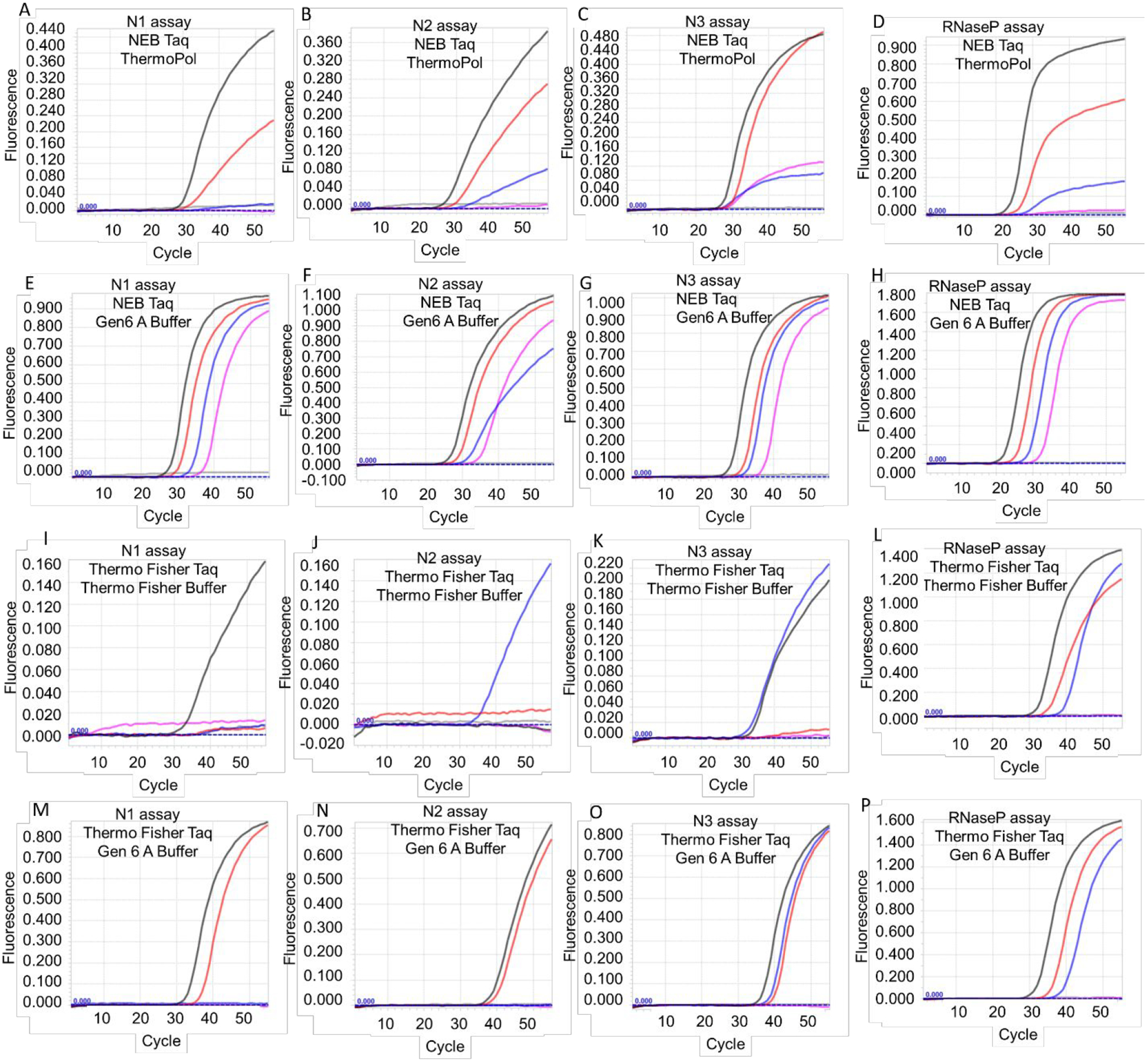
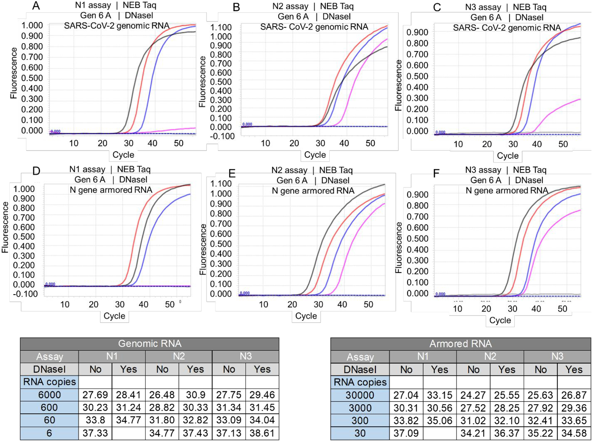
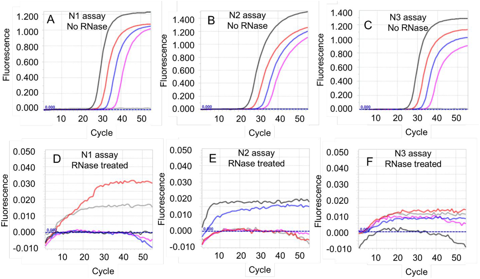
Taq DNA polymerase, one of the first thermostable DNA polymerases to be discovered, has been typecast as a DNA-dependent DNA polymerase commonly employed for PCR. However, Taq polymerase belongs to the same DNA polymerase superfamily as the Molony murine leukemia virus reverse transcriptase and has in the past been shown to possess reverse transcriptase activity. We report optimized buffer and salt compositions that promote the reverse transcriptase activity of Taq DNA polymerase and thereby allow it to be used as the sole enzyme in TaqMan RT-qPCRs. We demonstrate the utility of Taq-alone RT-qPCRs by executing CDC SARS-CoV-2 N1, N2, and N3 TaqMan RT-qPCR assays that could detect as few as 2 copies/μL of input viral genomic RNA.


Figures

Figure 1.
Figure 1.
SARS-CoV-2 N1 TaqMan RT-qPCR assays performed using NEB Taq DNA polymerase and N gene armored RNA in indicated buffers. Buffer compositions are detailed in Table 2. Representative amplification curves resulting from duplicate analysis of 3 × 10⁵ (black traces), 3 × 10⁴ (red traces), 3 × 10³ (blue traces), 3 × 10² (pink traces), 30 (green traces), and 0 (gray) copies of SARS-CoV-2 N gene armored RNA are depicted.
Figure 2.
Figure 2.
TaqMan RT-qPCR analysis of SARS-CoV-2 viral genomic RNA and RNaseP armored RNA using Taq DNA polymerase-based one-enzyme assays. CDC SARS-CoV-2 N gene assays, N1, N2, and N3, and RNaseP assay were performed using Taq DNA polymerase from either NEB (panels A-H) or Thermo Fisher (panels I-P). Assays were performed either using the companion commercial buffer (panels A-D and panels I-L) or using Gen 6 A buffer (panels E-H and panels M-P). Representative amplification curves from 6000 (black traces), 600 (red traces), 60 (blue traces), 6 (pink traces), and 0 (gray traces) copies of viral genomic RNA are depicted in panels A-C, E-G, I-K, and M-O. Representative amplification curves from 3 × 10⁵ (black traces), 3 × 10⁴ (red traces), 3 × 10³ (blue traces), 3 × 10² (pink traces) and 0 (gray traces) copies of armored RNaseP RNA are depicted in panes D, H, L, and P.
Figure 3.
Figure 3.
Effect of DNase I treatment on Taq DNA polymerase-mediated RT-qPCR assay. Taq DNA polymerase purchased from NEB was used to operate CDC SARS-CoV-2 N1, N2, and N3 TaqMan RT-qPCR assays using SARS-CoV-2 viral genomic RNA (panels A-C) or N gene armored RNA (panels D-F) treated with DNase I in duplicate experiments. Amplification curves shown in panels A-C resulted from 6000 (black traces), 600 (red traces), 60 (blue traces), 6 (pink traces), and 0 (gray traces) copies of SARS-CoV-2 genomic RNA. Amplification curves in panels D-F resulted from 30,000 (black traces), 3,000 (red traces), 300 (blue traces), 30 (pink traces) and 0 (gray traces) copies of N gene armored RNA. Representative Ct values for RT-qPCR amplification of indicated copies of untreated and DNase I treated SARS-CoV-2 genomic RNA and N gene armored RNA are tabulated.
Figure 4.
Figure 4.
Effect of RNase treatment on Taq DNA polymerase-mediated RT-qPCR assays. Taq DNA polymerase (NEB) was used to operate CDC SARS-CoV-2 N1, N2, and N3 TaqMan RT-qPCR assays using SARS-CoV-2 viral genomic RNA that had been pre-incubated either with zero RNase units (panels A-C) or with a combination of 1 unit of RNase A and 40 units of RNase T1 (panels D-F). Representative amplification curves from duplicate experiments using 6000 (black traces), 600 (red traces), 60 (blue traces), 6 (pink traces), and 0 (gray traces) copies of SARS-CoV-2 genomic RNA are depicted.

References

    1. Ellefson JW, Gollihar J, Shroff R, Shivram H, Iyer VR, and Ellington AD (2016) Synthetic evolutionary origin of a proofreading reverse transcriptase, Science 352, 1590–1593. - PubMed
    1. Loeb LA, Tartof KD, and Travaglini EC (1973) Copying natural RNAs with E. coli DNA polymerase I, Nat New Biol 242, 66–69. - PubMed
    1. Myers TW, and Gelfand DH (1991) Reverse transcription and DNA amplification by a Thermus thermophilus DNA polymerase, Biochemistry 30, 7661–7666. - PubMed
    1. Jackson LN, Chim N, Shi C, and Chaput JC (2019) Crystal structures of a natural DNA polymerase that functions as an XNA reverse transcriptase, Nucleic Acids Res 47, 6973–6983. - PMC - PubMed
    1. Jones MD, and Foulkes NS (1989) Reverse transcription of mRNA by Thermus aquaticus DNA polymerase, Nucleic acids research 17, 8387–8388. - PMC - PubMed