. 2021 Apr 1;29(4):320-329.e4.
doi: 10.1016/j.str.2020.11.011. Epub 2020 Dec 3.

Phosphorylation of RIAM by src promotes integrin activation by unmasking the PH domain of RIAM

Affiliations

Phosphorylation of RIAM by src promotes integrin activation by unmasking the PH domain of RIAM

Eun-Ah Cho et al. Structure. .

Abstract

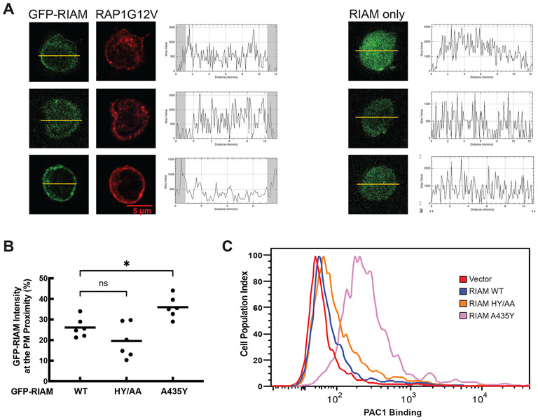
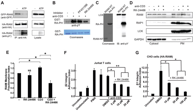
Integrin activation controls cell adhesion, migration, invasion, and extracellular matrix remodeling. RIAM (RAP1-GTP-interacting adaptor molecule) is recruited by activated RAP1 to the plasma membrane (PM) to mediate integrin activation via an inside-out signaling pathway. This process requires the association of the pleckstrin homology (PH) domain of RIAM with the membrane PIP2. We identify a conserved intermolecular interface that masks the PIP2-binding site in the PH domains of RIAM. Our data indicate that phosphorylation of RIAM by Src family kinases disrupts this PH-mediated interface, unmasks the membrane PIP2-binding site, and promotes integrin activation. We further demonstrate that this process requires phosphorylation of Tyr267 and Tyr427 in the RIAM PH domain by Src. Our data reveal an unorthodox regulatory mechanism of small GTPase effector proteins by phosphorylation-dependent PM association of the PH domain and provide new insights into the link between Src kinases and integrin signaling.

Keywords: FYN; LCK; PH domain; PIP2 binding; RAP1; RIAM; Src kinase; integrin signaling; lamellipodin; phosphorylation.


Conflict of interest statement

Declaration of interests The authors declare no competing interests.

Figures

Figure 1.
Figure 1.. Identifying a conserved intermolecular interface in RIAM.
A. Schematic representation of the domain organization of RIAM. Full length mouse RIAM (FL) possesses 670 residues. Talin-binding site (TBS) is colored in green; inhibitory (IN) segment is in orange; Coiled coil (CC) segment is in red; poly-proline (PP) segment is in black; the Ras-associating domain (RA) is in yellow; and the Pleckstrin-Homology domain (PH) is in cyan. The length of each structural region is proportional to the number of their amino acids. A scale bar corresponding to 100 amino acids is shown in gray. B. A conserved asymmetrical interface observed in the previously reported RIAM structures (3TCA: the original CC-RA-PHa; 4KVG: RIAM RA-PH in complex with RAP1-GTP; 6E31: RIAM in an autoinhibited configuration). One molecule was labeled as RA and PH, and the other molecule was labeled as RA’ and PH’. C. Left: superposition of the RIAM RA-PH core domain structure (wheat/pale cyan) and the new RIAM CC-RA-PHb structure (yellow/cyan). For each structure, two molecules (A and B) the form the asymmetrical interface are included in the superposition to demonstrate the identical interface (in dashed box). Right: conserved side-chain interactions that mediate the asymmetrical interface. Salt-bridge interactions are illustrated by yellow dash line; residues making Van der Waals contacts are indicated by surface representation. phosphoinositide-binding residues (K327, K328, K331, R332, and R333) are also indicated in stick representation. Ala435 is shown in the right panel to indicate its close contact with the neighboring molecule.
Figure 2.
Figure 2.. Mutation disrupting the interface enhances RIAM function.
A. Left: Jurkat T cells co-transfected with HA-tagged RAP1G12V and GFP-tagged RIAM. Cellular distribution of RIAM and RAP1 was plotted after densitometric analysis. Portions defined for the plasma membrane distribution are indicated by the shadowed areas in the plots. Right: Jurkat T cells transfected with GFP-RIAM alone. B. The PM distribution of RIAM (wild type and various mutations) is shown in the scatter plot. Statistical significances were calculated using a two-tailed t-test for unpaired samples. Additional cells used for analyzing PM distribution are shown in Fig. S2. C. αIIbβ3 over-expressed stable CHO cells (CHO-A5) were transfected with GFP or assorted GFP-RIAM constructs (WT, H389A/Y398A, and A435Y). The cells were incubated with PAC1 antibody (indicator of the integrin activation) and Alexa 647 to quantify GFP-tagged protein expression and integrin αIIbβ3 activity. Data were analyzed on a LSR flow cytometer using 30,000 cells per measurement.
Figure 3.
Figure 3.. RIAM phosphorylation by Src Promotes the PM translocation of RIAM and integrin activation.
A. Co-expression of HA-tagged RIAM and GFP-tagged RIAM in HEK293 cells. The cell lysate was treated with or without ATP, then subject to co-immunoprecipitation using anti-HA antibody, and immunoblotted with anti-GFP, anti-HA, and anti-pTyr antibodies. B. GST-RA-PH was treated with Jurkat T cell lysates (unstimulated or stimulated by anti-CD3), then pulled down by glutathione beads to examine for tyrosine phosphorylation. Lane 1: unstimulated; lane 2: stimulated; lane 3: stimulated and with 400 nM FAK inhibitor (PF431396); and lane 4: stimulated and with 400 nM Src kinase inhibitor (RK-24466). C. Purified RIAM-NT or RIAM RA-PH treated with LCK were examined for tyrosine phosphorylation. D. Jurkat T cells were starved and treated with RK-24466 and/or anti-CD3 antibodies. Cells were sub-fractionated and loaded for western blotting of cytosol and plasma membrane. A representative result of a triplicate experiment is shown. E. The PM distribution of RIAM indicated in 3D is shown in the bar graph. F. Jurkat T cells were starved and treated with RK-24466 and stimulated with or without anti-CD3 antibodies. Adhesion assay was performed and adherence was determined by plate reader (595 nm) with crystal violet. G. CHO cells were treated Src inhibitor for 30 min and adhesion assay was performed and adherence was determined by plate reader (595 nm) with crystal violet. E, F, and G: Data shown are mean ± SD, n = 3, also seen in Supplemental Table 1. Statistical significances were calculated using a two-tailed t-test for unpaired samples. *: p < 0.03; **: p < 0.005; #: p = 0.06 (NS).
Figure 4.
Figure 4.. Roles of Tyr267 and Tyr427 of RIAM in regulating the PM translocation of RIAM and integrin activation.
A. Four tyrosine phosphorylation sites (Tyr267, Tyr277, Tyr 398, and Tyr 427) in RIAM RA-PH (yellow and cyan) by LCK and Fyn kinases are indicated by sticks in green. A neighboring RA-PH molecule is shown in dark yellow (RA domain) and light blue (PH domain) with the PIP2 binding site highlighted in purple. B. Jurkat T cells were co-transfected with HA-tagged RAP1G12V and GFP-tagged RIAM. Cellular distribution of RIAM and RAP1 along the indicated bars was plotted after densitometric analysis. C. The PM distribution of RIAM (wild type and various mutations) is shown in the scatter plot. D. HEK293 cells co-transfected with HA-tagged RAP1G12V and GFP-tagged RIAM were sub-fractionated. Fractions of plasma membrane and cytosol were analyzed by Western blot. E. The PM distribution of RIAM indicated in 4D is shown in the bar graph. Data shown are mean ± SD, n = 3, also seen in Supplemental Table 1. F. CHO-A5 cells transfected with GFP-tagged RIAM were analyzed for αIIbβ3 integrin activity by FACS using PAC-1 antibody. Statistical significances were calculated using a two-tailed t-test for unpaired samples. *: p < 0.05.
Figure 5.
Figure 5.. A model of RIAM activation by FAK and Src kinases.
Dormant RIAM adopts an autoinhibited configuration with intermolecular interactions mediated by the PH domain and the N-terminal region. This autoinhibited configuration is released upon phosphorylation by FAK and Src kinases. FAK kinase phosphorylates the IN segment, unmasking the RAP1-binding site in the RA domain. Src kinase phosphorylates of the PH domain, unmasking the phosphoinositide-binding site in the PH domain. The two activating mechanisms of RA-PH by phosphorylation cooperatively activate RIAM, promoting the PM translocation of RIAM by interacting with RAP1 and PIP2.

Comment in

References

    1. Barczyk M, Carracedo S, and Gullberg D (2010). Integrins. Cell Tissue Res 339, 269–280. - PMC - PubMed
    1. Berlin-Rufenach C, Otto F, Mathies M, Westermann J, Owen MJ, Hamann A, and Hogg N (1999). Lymphocyte migration in lymphocyte function-associated antigen (LFA)-1-deficient mice. J Exp Med 189, 1467–1478. - PMC - PubMed
    1. Chang YC, Su W, Cho EA, Zhang H, Huang Q, Philips MR, and Wu J (2019). Molecular basis for autoinhibition of RIAM regulated by FAK in integrin activation. Proc Natl Acad Sci U S A 116, 3524–3529. - PMC - PubMed
    1. Chang YC, Zhang H, Franco-Barraza J, Brennan ML, Patel T, Cukierman E, and Wu J (2014). Structural and mechanistic insights into the recruitment of talin by RIAM in integrin signaling. Structure 22, 1810–1820. - PMC - PubMed
    1. De Franceschi N, Peuhu E, Parsons M, Rissanen S, Vattulainen I, Salmi M, Ivaska J, and Pouwels J (2015). Mutually Exclusive Roles of SHARPIN in Integrin Inactivation and NF-kappaB Signaling. PLoS One 10, e0143423. - PMC - PubMed

Publication types

MeSH terms

LinkOut - more resources