. 2021 Mar:144:115776.
doi: 10.1016/j.bone.2020.115776. Epub 2020 Dec 2.

A new approach to analyzing regenerated bone quality in the mouse digit amputation model using semi-automatic processing of microCT data

Affiliations

A new approach to analyzing regenerated bone quality in the mouse digit amputation model using semi-automatic processing of microCT data

Kevin F Hoffseth et al. Bone. 2021 Mar.

Abstract

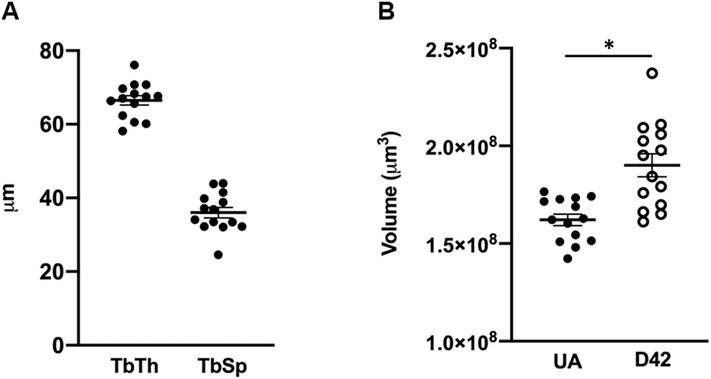
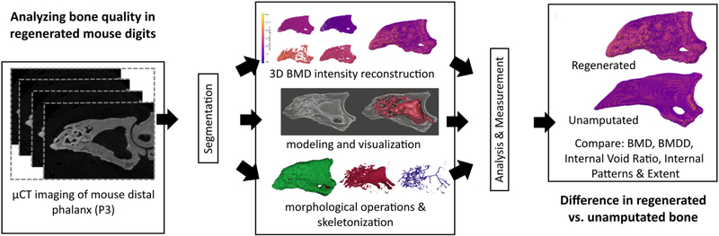
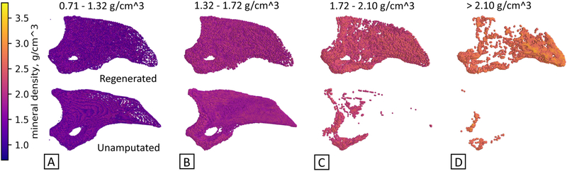
Bone regeneration is a critical area of research impacting treatment of diseases such as osteoporosis, age-related decline, and orthopaedic implants. A crucial question in bone regeneration is that of bone architectural quality, or how "good" is the regenerated bone tissue structurally? Current methods address typical long bone architecture, however there exists a need for improved ability to quantify structurally relevant parameters of bone in non-standard bone shapes. Here we present a new analysis approach based on open-source semi-automatic methods combining image processing, solid modeling, and numerical calculations to analyze bone tissue at a more granular level using μCT image data from a mouse digit model of bone regeneration. Examining interior architecture, growth patterning, spatial mineral content, and mineral density distribution, these methods are then applied to two types of 6-month old mouse digits - 1) those prior to amputation injury (unamputated) and 2) those 42 days after amputation when bone has regenerated. Results show regenerated digits exhibit increased inner void fraction, decreased patterning, different patterns of spatial mineral distribution, and increased mineral density values when compared to unamputated bone. Our approach demonstrates the utility of this new analysis technique in assessment of non-standard bone models, such as the regenerated bone of the digit, and aims to bring a deeper level of analysis with an open-source, integrative platform to the greater bone community.

Keywords: Bone pattern; Bone quality; Morphology; Regeneration; Semi-automatic; microCT.


Conflict of interest statement

Declaration of interest

Authors declare no competing interests.

Figures

Fig. A1.
Fig. A1.
Flowcharts of Spatial BMD and pre-skeletonization internal void processing methods. A) Creating 3D, spatial reconstructions of bone mineral density intensity and B) Process to reconstruct internal void volume prior to skeletonization.
Fig. 1.
Fig. 1.
Image processing based approach. Raw μCT images are acquired and subjected to segmentation, followed by 3D BMD reconstruction, interior modeling and visualization, and morphological mathematical operations including skeletonization. Following this, analysis and measurement evaluate BMD, BMDD, internal void ratio, and cumulative skeleton length.
Fig. 2.
Fig. 2.
Spatial visualization of mineral density values can illustrate differences in structure. Mineral density groups compare unamputated and regenerated digits across four mineral value ranges, A-D, with intensity scale at left. Regenerated digits show a significantly increased incidence of high mineral content as compared to the unamputated digit, in particular at the distal end. N = 3 UA, N = 3 D42. Representative images shown.
Fig. 3.
Fig. 3.
Mineral density value distribution. A) Single digit histograms compare individual unamputated (UA) and regenerated (D42) digits, showing the probability (frequency) and spread (density value range) of density values. Best fit curve with normal distribution. N = 1 UA, N = 1 D42. B) Right lateral view of full mineral density spectrum for both digits, showing distinctly contrasting spatial distribution of mineral density. N = 3 UA, N = 3 D42. Representative images shown.
Fig. 4.
Fig. 4.
Analysis of mineral density values for all unamputated and all regenerated digits shown as violin plots. Comparison of mineral content distribution between unamputated (UA) and regenerated (D42) digits. N = 1,559,938 values from N = 14 digits, UA. N = 858,333 values from N = 6 D42. Width of violin plot represents probability density.
Fig. 5.
Fig. 5.
Internal visualization of digits. A) Regenerated digit. B) Regenerated digit with internal void structure superimposed for clarity. C) Unamputated digit with internal void structure superimposed for clarity. Internal void structure is darkened for contrast, and colorized (red). N = 3. Representative images shown. (For interpretation of the references to color in this figure legend, the reader is referred to the web version of this article.)
Fig. 6.
Fig. 6.
Internal void space. A) Inner void fraction. Inner volume as a fraction of void space volume plus bone volume in unamputated (UA) and regenerated (D42) digits for distal volumetric region past the amputation plane. Mean = 0.0167 UA and 0.0913 D42, respectively. N = 14 UA. N = 6 D42. B) Total void space length. Results from measurement of skeletonized inner void space geometry in unamputated (UA) and regenerated (D42) digits also show an increase in internal void length in regenerated mice digits. S.D. = 256.1, and 1378.3, respectively. N = 14 UA. N = 6 D42. Skeletonization of the entire internal void space of digits to evaluate patterning, shown in C-E) regenerated and FH) unamputated representative digits. Whole bone is shown in green (C,F), inner void geometry shown in red (D,G), skeletonized void geometry shown in blue (E,H), for each representative sample shown (N = 3). (For interpretation of the references to color in this figure legend, the reader is referred to the web version of this article.)

References

    1. Lin X, Patil S, Gao YG, Qian A, The bone extracellular matrix in bone formation and regeneration, Front. Pharmacol. 11 (2020) 757. - PMC - PubMed
    1. Brockes JP, Kumar A, Appendage regeneration in adult vertebrates and implications for regenerative medicine, Science 310 (5756) (2005) 1919–1923. - PubMed
    1. Han M, Yang X, Lee J, Allan CH, Muneoka K, Development and regeneration of the neonatal digit tip in mice, Dev. Biol. 315 (1) (2008) 125–135. - PMC - PubMed
    1. Fernando WA, Leininger E, Simkin J, Li N, Malcom CA, Sathyamoorthi S, Han M, Muneoka K, Wound healing and blastema formation in regenerating digit tips of adult mice, Dev. Biol. 350 (2) (2011) 301–310. - PMC - PubMed
    1. Said S, Parke W, Neufeld DA, Vascular supplies differ in regenerating and nonregenerating amputated rodent digits, the anatomical record, Part A, Discoveries in Molecular, Cellular, and evolutionary Biology 278 (1) (2004) 443–449. - PubMed

Publication types

LinkOut - more resources