Review
. 2021 Apr 14;18(4):10.1088/1478-3975/abd0db.
doi: 10.1088/1478-3975/abd0db.

Roadmap for the multiscale coupling of biochemical and mechanical signals during development

Affiliations
Review

Roadmap for the multiscale coupling of biochemical and mechanical signals during development

Pierre-François Lenne et al. Phys Biol. .

Abstract

The way in which interactions between mechanics and biochemistry lead to the emergence of complex cell and tissue organization is an old question that has recently attracted renewed interest from biologists, physicists, mathematicians and computer scientists. Rapid advances in optical physics, microscopy and computational image analysis have greatly enhanced our ability to observe and quantify spatiotemporal patterns of signalling, force generation, deformation, and flow in living cells and tissues. Powerful new tools for genetic, biophysical and optogenetic manipulation are allowing us to perturb the underlying machinery that generates these patterns in increasingly sophisticated ways. Rapid advances in theory and computing have made it possible to construct predictive models that describe how cell and tissue organization and dynamics emerge from the local coupling of biochemistry and mechanics. Together, these advances have opened up a wealth of new opportunities to explore how mechanochemical patterning shapes organismal development. In this roadmap, we present a series of forward-looking case studies on mechanochemical patterning in development, written by scientists working at the interface between the physical and biological sciences, and covering a wide range of spatial and temporal scales, organisms, and modes of development. Together, these contributions highlight the many ways in which the dynamic coupling of mechanics and biochemistry shapes biological dynamics: from mechanoenzymes that sense force to tune their activity and motor output, to collectives of cells in tissues that flow and redistribute biochemical signals during development.

Keywords: embryogenesis; morphogenesis; signalling.


Figures

Figure 1.
Figure 1.
Illustration of a hypothetical morphogen-regulated developing tissue. Morphogen production by a restricted source (orange), spreading and degradation lead to the formation of a concentration gradient across the tissue. Target cells interpret this gradient and adopt red, green or blue cell identities. In this example, high morphogen concentrations promote cell proliferation and cell survival. As the tissue grows, morphogen levels in the expanding tissue are diluted by cell division, target cells move away from the source and the source itself also grows. The balance between these processes affects the final gradient shape, pattern and growth of the tissue.
Figure 2.
Figure 2.
Plant morphogenesis is a multiscale and multilevel problem.
Figure 3.
Figure 3.
(A) (Top) morphogen gradients form spatially varying concentration gradients across the system. (Middle) at threshold concentrations (dashed line), different downstream gene responses occur (bottom), defining spatial boundaries in the system. Reproduced from [18] under the CC BY 4.0 license. (B) Reaction–diffusion model for morphogen gradient formation (morphogen concentration denoted by ρ). SDD model, n = 1. (C) Steady-state profiles for the model shown in (B) for differing degradation terms, L = system length.
Figure 4.
Figure 4.
Possible interplay between tissue mechanics (A) and regulatory networks (B) to specify embryonic territories during quail gastrulation (C). Active forces are in magenta in (A); embryonic, mesendodermal, and extraembryonic territories are shown, respectively, in blue, red, and green, in (C).
Figure 5.
Figure 5.
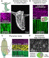
Parallel actin fibre arrays in different model systems. Images and schematics showing actin fibre arrays in (A) Hydra, (B) vertebrate gut (courtesy of Tyler Huycke, [53]), (C) planarian (courtesy of Lucila Scimone and Peter Reddien, [50]), and (D) Drosophila (courtesy of Maureen Cetera and Sally Horne-Badovinac, [52]).
Figure 6.
Figure 6.
Schematics of actin fibre array function and response to stress.
Figure 7.
Figure 7.
Schematics of possible couplings between geometry, mechanics (luminal pressure ΔP, apical constriction, cell stretching, fluid flows etc.) and biochemical signalling (receptor polarization, morphogen production/diffusion) that may drive supracellular patterning and morphogenesis. Art from Claudia Flandoli and modified from [59].
Figure 8.
Figure 8.
Tissue structure, defined as the composition, shape and three-dimensional arrangement of cells and extra-cellular matrices, arises through programs of self-organization. Tissue structure is linked to cell state through its impact on the microenvironment (green). The microenvironment is the sum of each paracrine signal impinging on a given cell, weighted by constraints provided by the local tissue structure. Active cell mechanics and other physical processes emerge from the molecular state of a cell and its ability to dissipate energy (red). Cell-generated forces result in shape changes and cell rearrangements that underlie many aspects of tissue-structure formation.
Figure 9.
Figure 9.
Experiments where tuning the levels of signalling molecules shifts patterns have been interpreted as evidence that the spatial gradient of signals sets up a molecular blueprint. What has not been taken into account is that such signals most likely modulate the physical behaviours of cells, which in turn changes the manner in which cells come together to construct the pattern.
Figure 10.
Figure 10.
Morphogenesis involves critical events across scales. Mechanical aspects of multicellular mechanics may serve as a bridge between molecular regulators and tissue-level phenotypes.
Figure 11.
Figure 11.
(A) Factors that affect the shape of MT asters in early embryos, and thus contribute to position and orient centrosomes and cell divisions through length-dependent MT forces. (B) An iterative cycle of cell shape influencing division position and division influencing subsequent cell shapes and thus division orientation in the next cycle.
Figure 12.
Figure 12.
Stages and mechanisms of control of cleavage divisions in early embryogenesis of Drosophila. (A) Nuclear-cycle stages. Following fertilization, the nuclear cycles are characterized by stereotypical movements: axial expansion denotes the movement of nuclei along the AP axis (cycles 4–6); cortical migration denotes the movement of nuclei to the cortex (cycle 7–9). (B) Model for axial expansion as in proposed in [120].
Figure 13.
Figure 13.
Examples where contact morphology affects cell fate. (A) In a chick’s inner ear, smaller cells are more likely to become hair cells [127]. (B) Fate switch in the Drosophila intestine depends on contact area [128]. (C) Signalling level is weaker for cells contacting through basal filopodia compared with cells in direct apical contact in Drosophila bristle patterning [129].
Figure 14.
Figure 14.
Dependence of Notch signalling on contact area. (A) Schematic of Notch signalling. (B) Dependence of Notch signalling on contact width. Diffusion-limited regime occurs when the contact width is smaller than the diffusion-length scale. (C) Schematic of factors affecting contact dependence of signalling.
Figure 15.
Figure 15.
(A) Local tissue contractions can produce deformation/tissue flows over a length scale λ which is dependent on material properties. In the case of a viscous tissue in interaction with an external substrate, this length scale varies with tissue viscosity and the effective friction coefficient between the tissue and the substrate. (B) Geometric boundaries guide flow patterns. Simulations of tissue flows during Drosophila morphogenesis [138] demonstrate how a local tissue contraction in the germband can generate a global tissue flow whose geometry is influenced by the embryo’s geometry. In the absence of the cephalic furrow, which is a highly curved interface separating the anterior part and the posterior part of the embryo, the global tissue flow is highly affected. (C) Coupling between the tissue’s moving boundaries and bulk tissue flows in an organoid system. Mechanisms coupling the tissue boundary geometry (here, its curvature) with the generation of local active stresses can generate instabilities leading to the shape variability observed in organoid systems. Local mechanics and global geometry are coupled to give rise to shape: this coupling could be perturbed by imposing geometrical constraints on the organoid using micro-patterning, for example.

References

    1. Heemskerk I. Full of potential: pluripotent stem cells for the systems biology of embryonic patterning. Dev. Biol. 2019;460:86. - PubMed
    1. Shahbazi MN, Siggia ED and Zernicka-Goetz M 2019. Self-organization of stem cells into embryos: a window on early mammalian development Science 364 948–51 - PMC - PubMed
    1. Chhabra S, Liu L, Goh R, Kong X and Warmflash A 2019. Dissecting the dynamics of signaling events in the BMP, WNT, and NODAL cascade during self-organized fate patterning in human gastruloids PLoS Biol. 17 e3000498. - PMC - PubMed
    1. Britton G, Heemskerk I, Hodge R, Qutub AA and Warmflash A 2019. A novel self-organizing embryonic stem cell system reveals signaling logic underlying the patterning of human ectoderm Development 146 dev179093. - PMC - PubMed
    1. Haremaki T, Metzger JJ, Rito T, Ozair MZ, Etoc F and Brivanlou AH 2019. Self-organizing neuruloids model developmental aspects of Huntington’s disease in the ectodermal compartment Nat. Biotechnol 37 1198–208 - PubMed

Publication types

LinkOut - more resources