

Regenerative capacity of the corneal transition zone for endothelial cell therapy
- PMID: 33276809
- PMCID: PMC7716425
- DOI: 10.1186/s13287-020-02046-2
Regenerative capacity of the corneal transition zone for endothelial cell therapy
Abstract
The corneal endothelium located on the posterior corneal surface is responsible for regulating stromal hydration. This is contributed by a monolayer of corneal endothelial cells (CECs), which are metabolically active in a continuous fluid-coupled efflux of ions from the corneal stroma into the aqueous humor, preventing stromal over-hydration and preserving the orderly arrangement of stromal collagen fibrils, which is essential for corneal transparency. Mature CECs do not have regenerative capacity and cell loss due to aging and diseases results in irreversible stromal edema and a loss of corneal clarity. The current gold standard of treatment for this worldwide blindness caused by corneal endothelial failure is the corneal transplantation using cadaveric donor corneas. The top indication is Fuchs corneal endothelial dystrophy/degeneration, which represents 39% of all corneal transplants performed. However, the global shortage of transplantable donor corneas has restricted the treatment outcomes, hence instigating a need to research for alternative therapies. One such avenue is the CEC regeneration from endothelial progenitors, which have been identified in the peripheral endothelium and the adjacent transition zone. This review examines the evidence supporting the existence of endothelial progenitors in the posterior limbus and summarizes the existing knowledge on the microanatomy of the transitional zone. We give an overview of the isolation and ex vivo propagation of human endothelial progenitors in the transition zone, and their growth and differentiation capacity to the corneal endothelium. Transplanting these bioengineered constructs into in vivo models of corneal endothelial degeneration will prove the efficacy and viability, and the long-term maintenance of functional endothelium. This will develop a novel regenerative therapy for the management of corneal endothelial diseases.
Keywords: Cornea endothelium; Corneal endothelial progenitors; Schwalbe’s line, transitional zone, corneal endothelial cell degeneration.
Conflict of interest statement
No financial and non-financial competing interest exists for any author.
Figures

Similar articles
-
Specular Microscopy.2024 Apr 21. In: StatPearls [Internet]. Treasure Island (FL): StatPearls Publishing; 2025 Jan–. 2024 Apr 21. In: StatPearls [Internet]. Treasure Island (FL): StatPearls Publishing; 2025 Jan–. PMID: 36256774 Free Books & Documents.
-
Stem cell markers in the human posterior limbus and corneal endothelium of unwounded and wounded corneas.Mol Vis. 2007 Oct 18;13:1984-2000. Mol Vis. 2007. PMID: 17982423
-
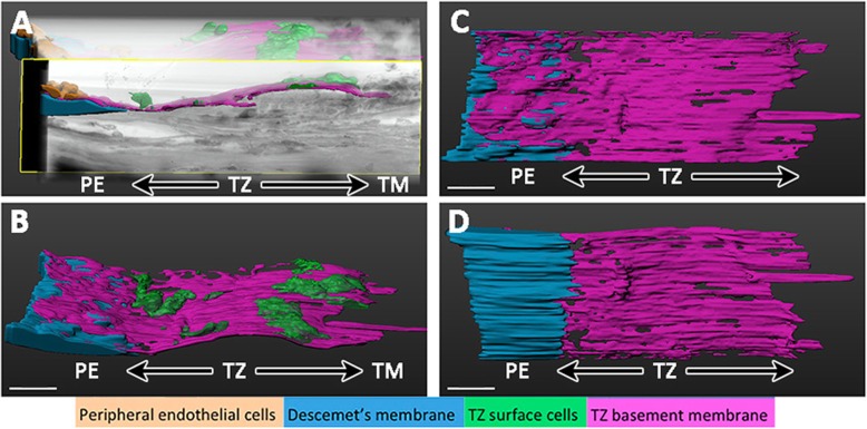
Characterization of Human Transition Zone Reveals a Putative Progenitor-Enriched Niche of Corneal Endothelium.Cells. 2019 Oct 12;8(10):1244. doi: 10.3390/cells8101244. Cells. 2019. PMID: 31614883 Free PMC article.
-
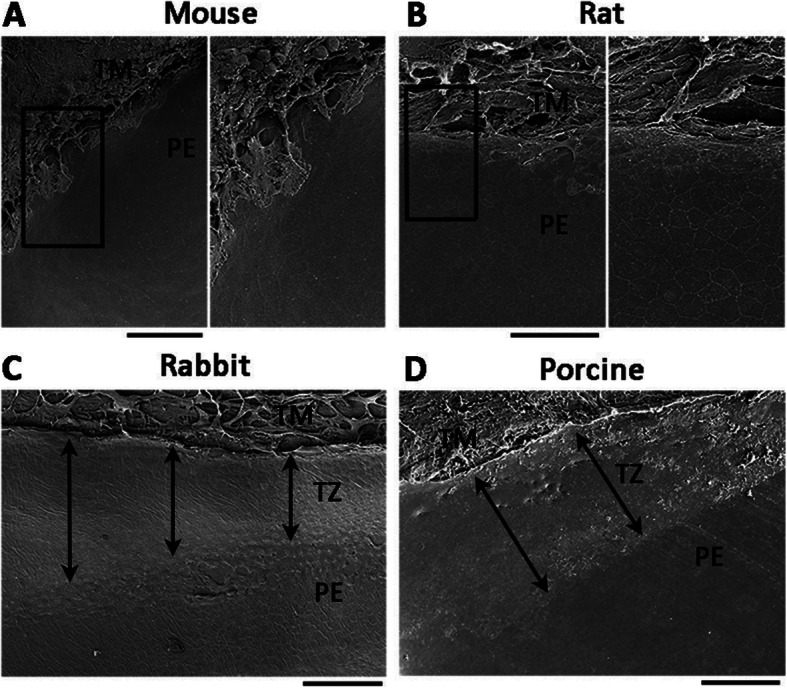
Proliferative capacity of the corneal endothelium.Prog Retin Eye Res. 2003 May;22(3):359-89. doi: 10.1016/s1350-9462(02)00065-4. Prog Retin Eye Res. 2003. PMID: 12852491 Review.
-
[Transplantation of corneal endothelial cells].Nippon Ganka Gakkai Zasshi. 2002 Dec;106(12):805-35; discussion 836. Nippon Ganka Gakkai Zasshi. 2002. PMID: 12610838 Review. Japanese.
Cited by
-
Biomedical Application of MSCs in Corneal Regeneration and Repair.Int J Mol Sci. 2025 Jan 15;26(2):695. doi: 10.3390/ijms26020695. Int J Mol Sci. 2025. PMID: 39859409 Free PMC article. Review.
-
Biofabrication of Artificial Stem Cell Niches in the Anterior Ocular Segment.Bioengineering (Basel). 2021 Sep 30;8(10):135. doi: 10.3390/bioengineering8100135. Bioengineering (Basel). 2021. PMID: 34677208 Free PMC article. Review.
-
The Interplay Between Metabolites and MicroRNAs in Aqueous Humor to Coordinate Corneal Endothelium Integrity.Ophthalmol Sci. 2023 Mar 16;3(3):100299. doi: 10.1016/j.xops.2023.100299. eCollection 2023 Sep. Ophthalmol Sci. 2023. PMID: 37125267 Free PMC article.
-
A Review of Corneal Blindness: Causes and Management.Cureus. 2022 Oct 9;14(10):e30097. doi: 10.7759/cureus.30097. eCollection 2022 Oct. Cureus. 2022. PMID: 36381769 Free PMC article. Review.
-
Assessing Corneal Endothelial Damage Using Terahertz Time-Domain Spectroscopy and Support Vector Machines.Sensors (Basel). 2022 Nov 23;22(23):9071. doi: 10.3390/s22239071. Sensors (Basel). 2022. PMID: 36501773 Free PMC article.
References
Publication types
MeSH terms
LinkOut - more resources
Full Text Sources
