. 2021 Jan-Jun:296:100149.
doi: 10.1074/jbc.RA120.016423. Epub 2020 Dec 10.

PIP2 promotes conformation-specific dimerization of the EphA2 membrane region

Affiliations

PIP2 promotes conformation-specific dimerization of the EphA2 membrane region

Katherine M Stefanski et al. J Biol Chem. 2021 Jan-Jun.

Abstract

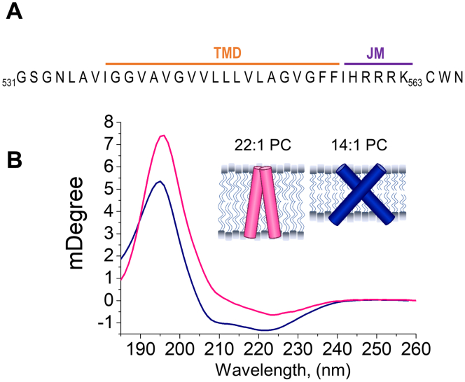
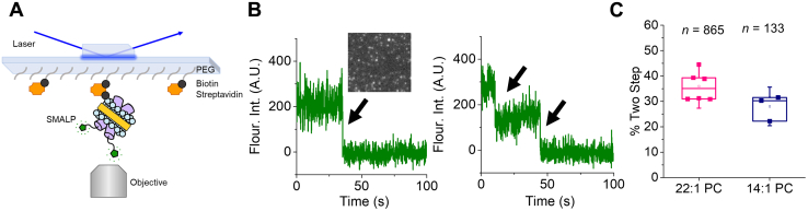
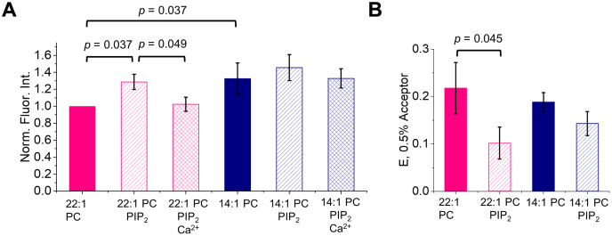
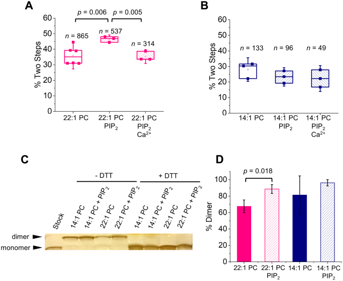
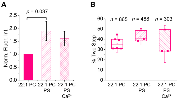
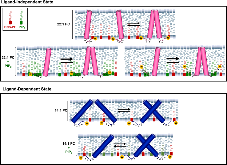
The impact of the EphA2 receptor on cancer malignancy hinges on the two different ways it can be activated. EphA2 induces antioncogenic signaling after ligand binding, but ligand-independent activation of EphA2 is pro-oncogenic. It is believed that the transmembrane (TM) domain of EphA2 adopts two alternate conformations in the ligand-dependent and the ligand-independent states. However, it is poorly understood how the difference in TM helical crossing angles found in the two conformations impacts the activity and regulation of EphA2. We devised a method that uses hydrophobic matching to stabilize two conformations of a peptide comprising the EphA2 TM domain and a portion of the intracellular juxtamembrane (JM) segment. The two conformations exhibit different TM crossing angles, resembling the ligand-dependent and ligand-independent states. We developed a single-molecule technique using styrene maleic acid lipid particles to measure dimerization in membranes. We observed that the signaling lipid PIP2 promotes TM dimerization, but only in the small crossing angle state, which we propose corresponds to the ligand-independent conformation. In this state the two TMs are almost parallel, and the positively charged JM segments are expected to be close to each other, causing electrostatic repulsion. The mechanism PIP2 uses to promote dimerization might involve alleviating this repulsion due to its high density of negative charges. Our data reveal a conformational coupling between the TM and JM regions and suggest that PIP2 might directly exert a regulatory effect on EphA2 activation in cells that is specific to the ligand-independent conformation of the receptor.

Keywords: AKT; SMALP; juxtamembrane; receptor tyrosine kinase; single-molecule; transmembrane.


Conflict of interest statement

Conflict of interest The authors declare that they have no conflicts of interest with the contents of this article.

Figures

Figure 1
Figure 1
Bilayer thickness drives differences in TMJM helical tilt.A, sequence of the TMJM peptide comprised of EphA2 residues 531 to 563 with added CWN residues at the C terminus. B, OCD spectra of TMJM in 22:1 PC (fuchsia) and 14:1 PC (navy). Inset, model of the conformations of TMJM dimer in 14:1 PC and 22:1 based on OCD data.
Figure 2
Figure 2
TMJM dimerization observed by single-molecule TIRF of SMALPs.A, schematic of TIRF experimental setup. SMALP showing SMA polymer (yellow) encircling lipids (blue) containing TMJM peptides (purple) labeled with Alexa Fluor 488 (green), immobilized on a PEGylated slide via a biotin (black) and streptavidin (orange) linkage. B, representative fluorescence traces showing one (left) and two (right) photobleaching steps (black arrows). Representative TIRF image of SMALPs (inset). C, box and whiskers plot (upper quartile, median, and lower quartile) showing percentages of peptide counted in traces with two photobleaching steps in 22:1 PC and 14:1 PC at a lipid-to-peptide ratio of 300:1. Data are from three to six independent experiments ±S.D, n is total number of traces counted with two photobleaching steps.
Figure 3
Figure 3
Bilayer thickness and PIP2alter Trp environment while PIP2changes headgroup distance.A, normalized intensities (350 nm) of TMJM Trp in 22:1 PC and 14:1 PC liposomes in the presence and absence of PIP2 and 5 mM Ca2+. p-values were determined by Mann–Whitney U tests, bars are means ± S.D., n = 3. B, FRET efficiencies calculated from FRET experiments with TMJM (Trp, donor) in 14:1 PC and 22:1 PC liposomes in the presence and absence of 3% PIP2 (0.5% DNS-PE, acceptor) in liposomes. Bars are means ± S.D., n = 3. p-value is from one-way ANOVA followed by Tukey post-hoc test.
Figure 4
Figure 4
PIP2promotes self-assembly of TMJM in thick bilayers.AB, percentage of Alexa Fluor 488 labeled TMJM peptide in two-step photobleaching traces in 22:1 PC and 14:1 SMALPs, respectively, determined by single-molecule studies. The effect of the presence of 3% PIP2 and 1 mM Ca2+ is investigated. Data are from three to six independent experiments. n is number traces counted for each. p-values are from Student’s t-tests with significance determined after Benjamini–Hochberg procedure. C, representative SDS-PAGE (full gel can be seen in Fig. S10) of unlabeled TMJM in 14:1 PC and 22:1 PC liposomes in the presence and absence of 3% PIP2. Monomer and disulfide-mediated dimers can be seen in nonreducing conditions. Addition of 5 mM DTT eliminates the disulfide-mediated dimer band. D, quantification of three independent SDS-PAGE experiments as shown in C. Bands in each lane were quantified in ImageJ and percent of dimer was calculated. Bars are means ± S.D., p-value is from a Student’s t-test. All data are from experiments at a lipid-to-peptide ratio of 300:1.
Figure 5
Figure 5
PS interactions with TMJM in thick bilayers.A, normalized fluorescence intensities from emission spectra of TMJM in 22:1 PC liposomes with 10% POPS and 5 mM Ca2+. Bars are means ± SD, n = 3. p-value was determined by Mann–Whitney U test. B, percentages of dimeric TMJM from SM-photobleaching experiments in 22:1 PC examining effects of 10% POPS and 1 mM Ca2+. Data are from three to six independent experiments. n is number traces counted for each. No statistically significant differences were found.
Figure 6
Figure 6
Model TMJM showing different effect of PIP2on two TMJM configurations. Top, in the ligand-independent signaling configuration TMJM exists in a monomer–dimer equilibrium in the absence of PIP2. Charge–charge repulsion of the JMs must be overcome for efficient dimerization. In the presence of PIP2 the JM–lipid association changes by either clustering of PIP2 around the JM or burial of the JM. This shields the positive charges promoting dimerization. Bottom, in the ligand-independent signaling configuration, due to the tilt of the TM, charge repulsion of the JMs is not present. Neither JM environment nor dimerization is altered by PIP2.

References

    1. Boyd A.W., Bartlett P.F., Lackmann M. Therapeutic targeting of EPH receptors and their ligands. Nat. Rev. Drug Discov. 2014;13:39–62. - PubMed
    1. Pasquale E.B. Eph-Ephrin bidirectional signaling in physiology and disease. Cell. 2008;133:38–52. - PubMed
    1. Fu A.K.Y., Hung K.-W., Huang H., Gu S., Shen Y., Cheng E.Y.L., Ip F.C.F., Huang X., Fu W.-Y., Ip N.Y. Blockade of EphA4 signaling ameliorates hippocampal synaptic dysfunctions in mouse models of Alzheimer’s disease. Proc. Natl. Acad. Sci. U. S. A. 2014;111:9959–9964. - PMC - PubMed
    1. Vargas L.M., Leal N., Estrada L.D., González A., Serrano F., Araya K., Gysling K., Inestrosa N.C., Pasquale E.B., Alvarez A.R. EphA4 activation of c-Abl mediates synaptic loss and LTP blockade caused by amyloid-β oligomers. PLoS One. 2014;9 - PMC - PubMed
    1. Van Hoecke A., Schoonaert L., Lemmens R., Timmers M., Staats K.A., Laird A.S., Peeters E., Philips T., Goris A., Dubois B., Andersen P.M., Al-Chalabi A., Thijs V., Turnley A.M., Van Vught P.W. EPHA4 is a disease modifier of amyotrophic lateral sclerosis in animal models and in humans. Nat. Med. 2012;18:1418–1422. - PubMed

Publication types

Substances

LinkOut - more resources