. 2020 Dec 4;5(1):283.
doi: 10.1038/s41392-020-00426-x.

CD147-spike protein is a novel route for SARS-CoV-2 infection to host cells

Affiliations

CD147-spike protein is a novel route for SARS-CoV-2 infection to host cells

Ke Wang et al. Signal Transduct Target Ther. .

Abstract

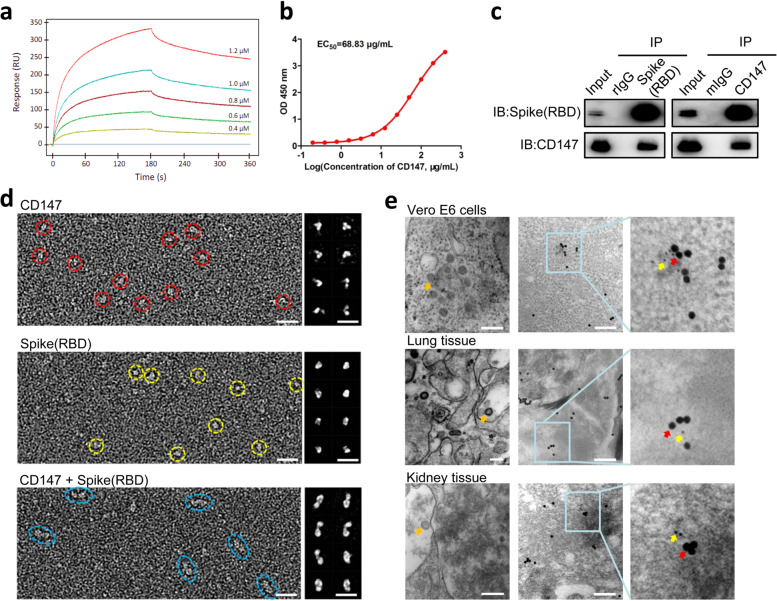
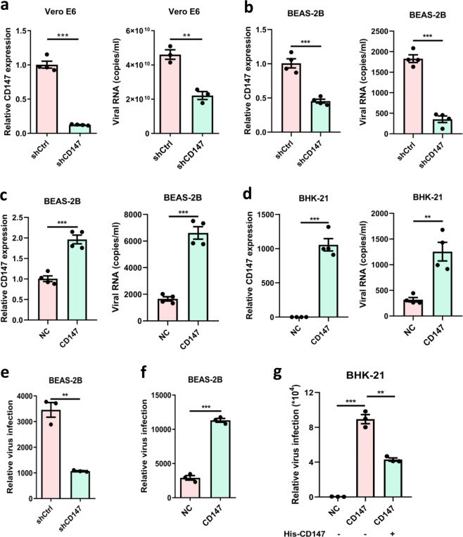
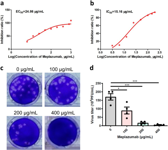
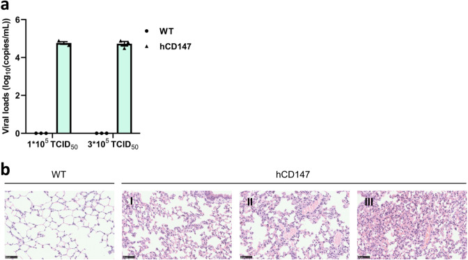
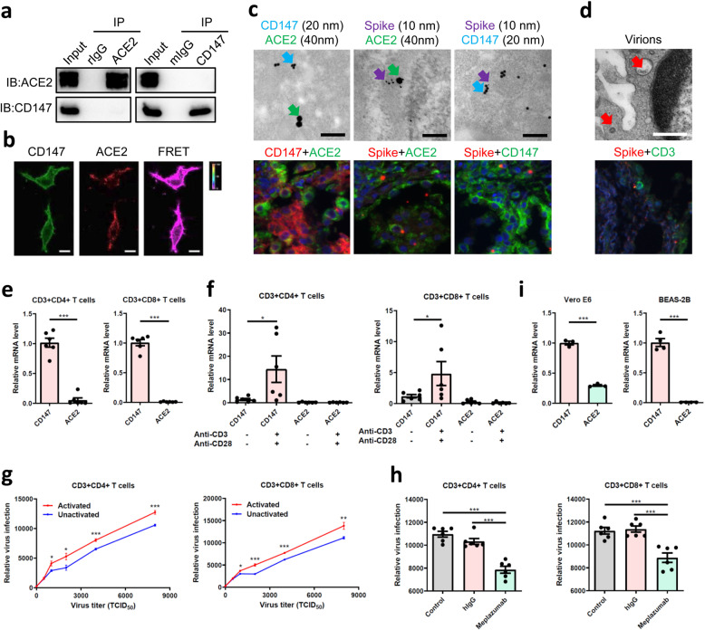
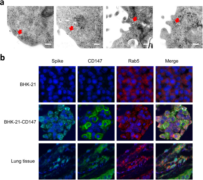
In face of the everlasting battle toward COVID-19 and the rapid evolution of SARS-CoV-2, no specific and effective drugs for treating this disease have been reported until today. Angiotensin-converting enzyme 2 (ACE2), a receptor of SARS-CoV-2, mediates the virus infection by binding to spike protein. Although ACE2 is expressed in the lung, kidney, and intestine, its expressing levels are rather low, especially in the lung. Considering the great infectivity of COVID-19, we speculate that SARS-CoV-2 may depend on other routes to facilitate its infection. Here, we first discover an interaction between host cell receptor CD147 and SARS-CoV-2 spike protein. The loss of CD147 or blocking CD147 in Vero E6 and BEAS-2B cell lines by anti-CD147 antibody, Meplazumab, inhibits SARS-CoV-2 amplification. Expression of human CD147 allows virus entry into non-susceptible BHK-21 cells, which can be neutralized by CD147 extracellular fragment. Viral loads are detectable in the lungs of human CD147 (hCD147) mice infected with SARS-CoV-2, but not in those of virus-infected wild type mice. Interestingly, virions are observed in lymphocytes of lung tissue from a COVID-19 patient. Human T cells with a property of ACE2 natural deficiency can be infected with SARS-CoV-2 pseudovirus in a dose-dependent manner, which is specifically inhibited by Meplazumab. Furthermore, CD147 mediates virus entering host cells by endocytosis. Together, our study reveals a novel virus entry route, CD147-spike protein, which provides an important target for developing specific and effective drug against COVID-19.


Conflict of interest statement

The authors declare no competing interests.

Figures

Fig. 1
Fig. 1
Identification of the interaction and co-localization between CD147 and spike protein. ac The interaction of CD147 and spike was detected by SPR assay (a), ELISA (b), and Co-IP assay (c). The mouse IgG (mIgG) and rabbit IgG (rIgG) were served as negative controls. d OpNS-EM images of CD147, spike(RBD) and CD147-spike(RBD) complexes. Left panels showed the survey view of the micrograph. Right panels showed 8 class averages were selected from a total of more than 300 class averages which were respectively calculated from a total of 6,681 CD147 particles; 5,073 particles of spike(RBD); 12,426 particles of CD147-spike(RBD) complexes. Scale bars: 10 nm. e The co-localization of CD147 and spike protein was observed by immuno-electron microscope. Virions (orange arrows) were observed in virus-infected Vero E6 cells and lung and kidney tissues from COVID-19 patient. The co-localization of CD147 (20 nm-gold colloid, red arrows) and spike protein (10 nm-gold colloid, yellow arrows) in SARS-CoV-2 infected Vero E6 cells and lung and kidney tissues from a patient with COVID-19. Scale bars: 200 nm
Fig. 2
Fig. 2
SARS-CoV-2 employs the CD147 receptor for host cell entry. a, b Left, Vero E6 and BEAS-2B cells were transfected with shRNA for CD147 gene silence; the gene expression level was detected by real-time PCR. Right, SARS-CoV-2 infection test was performed in Vero E6-shCD147 and BEAS-2B-shCD147 cells. At 48 h after infection, the virus copy number was detected with quantitative PCR (**p < 0.01, ***p < 0.001, two-tailed t-test, mean ± SEM). c, d Left, CD147 expression level was detected by real-time PCR in BEAS-2B-CD147 and BHK-21-CD147 cells. Right, SARS-CoV-2 infection test was performed in BEAS-2B-CD147 and BHK-21-CD147 cells. At 48 h after infection, the virus copy number was detected with quantitative PCR (**p < 0.01, ***p < 0.001, two-tailed t-test, mean ± SEM). e, f The infection efficiency of SARS-CoV-2 pseudovirus was detected by luciferase reporter assay in BEAS-2B-shCD147 and BEAS-2B-CD147 cells (**p < 0.01, ***p < 0.001, two-tailed t-test, mean ± SEM). g SARS-CoV-2 pseudovirus infection of BHK-21-CD147 cells was neutralized by recombinant human CD147 extracellular fragment (**p < 0.01, ***p < 0.001, two-tailed t-test, mean ± SEM)
Fig. 3
Fig. 3
Meplazumab significantly inhibited virus replication. a, b Virus inhibition assays were performed to calculate EC50 and IC50 by crystal violet staining and quantitative PCR, respectively. c, d The inhibitory effect of Meplazumab on virus infection was detected by plaque assay and virus titer was quantified (*p < 0.05, ***p < 0.001, two-tailed t-test, mean ± SEM)
Fig. 4
Fig. 4
SARS-CoV-2 invades lung tissues of hCD147 mice and causes pathologic changes. a The WT mice and hCD147 mice were infected with SARS-CoV-2. At 48 h after infection, the viral load of lung tissues was detected with quantitative PCR. b The histopathological changes of lung tissues were detected in WT mice and hCD147 mice by HE staining. Scale bars: 50 μm
Fig. 5
Fig. 5
CD147 is an alternative receptor for SARS-CoV-2 infection in ACE2-deficient cell types. a No interaction of CD147 and ACE2 was detected by Co-IP assay. The mIgG and rIgG were served as negative controls. b No co-localization was found between CD147 and ACE2 by FRET. The color bar denotes FRET ratio. Scale bars: 10 μm. c No co-localization of CD147-ACE2 and the co-localizations of spike-ACE2 and spike-CD147 were observed by immuno-electron microscope (scale bars: 200 nm) and multicolor immunofluorescence (magnification: ×200) in lung tissues from COVID-19 patient. Spike protein, 10 nm-gold colloid, purple arrows; CD147, 20 nm-gold colloid, blue arrows; and ACE2, 40 nm-gold colloid, green arrows. d Virions (red arrows) were observed in lymphocytes of lung tissues from COVID-19 patient. Scale bars: 500 nm. The localization of spike protein and CD3 was analyzed by multicolor immunofluorescence staining. Magnification: ×200. e The gene expressions of CD147 and ACE2 in CD4+ and CD8+ T cells were detected by real-time PCR (***p < 0.001, two-tailed t-test, mean ± SEM). f The expressions of CD147 and ACE2 in unactivated or activated T cells were detected by real-time PCR (*p < 0.05, Mann–Whitney test, mean ± SEM). g The infection efficiency of SARS-CoV-2 pseudovirus was detected by luciferase reporter assay in unactivated and activated T cells (*p < 0.05, **p < 0.01, ***p < 0.001, two-tailed t-test, mean ± SEM). h SARS-CoV-2 pseudovirus infection of CD4+ and CD8+ T cells was inhibited by Meplazumab (***p < 0.001, two-tailed t-test, mean ± SEM). i The gene expressions of CD147 and ACE2 in Vero E6 and BEAS-2B cells were detected by real-time PCR (***p < 0.001, two-tailed t-test, mean ± SEM)
Fig. 6
Fig. 6
SARS-CoV-2 enters the host cells through CD147-mediated endocytosis. a The sequential endocytosis of SARS-CoV-2 was observed in Vero E6 cells by electron microscope. Scale bars: 200 nm. b The co-localization of spike protein, CD147, and Rab5 were analyzed in BHK-21-CD147 cells and lung tissues from COVID-19 patient by multicolor immunofluorescence staining. Magnification: ×200.

References

    1. Jiang S, Xia S, Ying T, Lu L. A novel coronavirus (2019-nCoV) causing pneumonia-associated respiratory syndrome. Cell. Mol. Immunol. 2020;5:554. doi: 10.1038/s41423-020-0372-4. - DOI - PMC - PubMed
    1. Liu K, et al. Clinical characteristics of novel coronavirus cases in tertiary hospitals in Hubei Province. Chin. Med. J. 2020;9:1025–1031. doi: 10.1097/CM9.0000000000000744. - DOI - PMC - PubMed
    1. Xu Z, et al. Pathological findings of COVID-19 associated with acute respiratory distress syndrome. Lancet. Respir. Med. 2020;4:420–422. doi: 10.1016/S2213-2600(20)30076-X. - DOI - PMC - PubMed
    1. Zhang, L. et al. The D614G mutation in the SARS-CoV-2 spike protein reduces S1 shedding and increases infectivity. Preprint at https://www.ncbi.nlm.nih.gov/pmc/articles/PMC7310631/ (2020).
    1. Hulswit RJ, de Haan CA, Bosch BJ. Coronavirus spike protein and tropism changes. Adv. Virus Res. 2016;96:29–57. doi: 10.1016/bs.aivir.2016.08.004. - DOI - PMC - PubMed

Publication types

MeSH terms

LinkOut - more resources