. 2020 Dec 4;10(1):21295.
doi: 10.1038/s41598-020-78304-3.

Coiled-coil domain containing 50-V2 protein positively regulates neurite outgrowth

Affiliations

Coiled-coil domain containing 50-V2 protein positively regulates neurite outgrowth

Ju-Sik Min et al. Sci Rep. .

Abstract

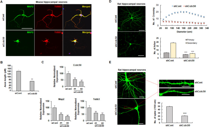
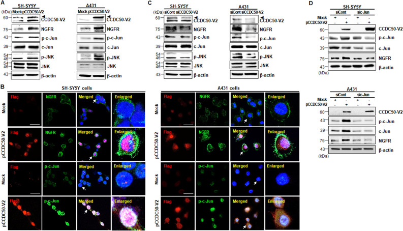
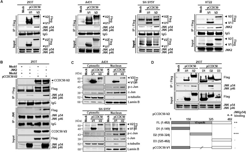
The coiled-coil domain containing 50 (CCDC50) protein is a phosphotyrosine-dependent signalling protein stimulated by epidermal growth factor. It is highly expressed in neuronal cells in the central nervous system; however, the roles of CCDC50 in neuronal development are largely unknown. In this study, we showed that the depletion of CCDC50-V2 impeded the neuronal development process, including arbor formation, spine density development, and axonal outgrowth, in primary neurons. Mechanistic studies revealed that CCDC50-V2 positively regulated the nerve growth factor receptor, while it downregulated the epidermal growth factor receptor pathway. Importantly, JNK/c-Jun activation was found to be induced by the CCDC50-V2 overexpression, in which the interaction between CCDC50-V2 and JNK2 was also observed. Overall, the present study demonstrates a novel mechanism of CCDC50 function in neuronal development and provides new insight into the link between CCDC50 function and the aetiology of neurological disorders.


Conflict of interest statement

The authors declare no competing interests.

Figures

Figure 1
Figure 1
CCDC50 knockdown suppresses dendritic and axonal outgrowth in primary cultured neurons. (A-C) Mouse primary hippocampal neurons were transiently transfected with control siRNA (siCont) and siRNAs against the murine CCDC50 gene on day in vitro 3 (DIV 3). (A) After 72 h of incubation, the cells were stained for MAP2 (green) and TUBB3 (red) (A). Scale bars = 30 µm. (B) Axonal lengths were measured and are presented as the proportion of the dendritic length of TUBB3-positive and MAP2-negative dendrites. Over 40 cells were measured in three independent experiments. (C) The expression levels of neuronal markers such as Map2 and Tubb3 in primary neurons treated with two siRNAs against Ccdc50 (siCcdc50# 1 and siCcdc50# 2) were analysed by quantitative real time RT-PCR. Data presented were normalized to Gapdh. (D, E) Analysis of the attenuation of dendritic arbor development and spine density in Ccdc50 knockdown neuronal cells. Rat primary hippocampal neurons were stably transfected with green fluorescent protein (GFP)-tagged control (shCont) or shCcdc50 (a vector that silences all variants of rat Ccdc50) on DIV 5; then, immunostaining with an anti-EGFP antibody was performed on DIV 9 (D; left panel). The quantification of dendrite complexity by Sholl analysis of rat primary hippocampal neurons (D; upper right). The numbers of primary and secondary dendrites were lower in Ccdc50 knockdown cells than in control cells (D; lower right). Means ± SD of data from 30 control neurons and 30 CCDC50 knock down neurons are shown. (E) The densities of the dendritic spines were compared between control (shCont)- and shCcdc50-treated cells. Means ± SD of data from 84 dendrites for control and 79 dendrites for CCDC50 knock down are shown. *p < 0.05, **p < 0.01, ***p < 0.001, Student’s t-test compared with the control group.
Figure 2
Figure 2
CCDC50-V2 knockdown decreases neuronal differentiation. (A) Human SH-SY5Y neuroblastoma cells were transfected with shCont, shCCDC50-V1 (human CCDC50-V1) and shCCDC50-V2 (human CCDC50-V2) and then treated with retinoic acid (RA, 10 µM) for the indicated time periods. The cells were immunostained with anti-EGFP (green), anti-TUBB3 (red, neuron-specific actin cytoskeleton), and DAPI (blue, nuclei). Scale bar = 100 µm. Neurite lengths were measured in approximately 30 GFP-expressing cells and compared in three independent experiments (A; lower panel) and graphically presented. (B) The expression levels of MAP2 and TUBB3 in transfected cells were measured using western blotting (upper panels) and semi-quantitative RT-PCR analysis (lower panels). (C) The mRNA expression patterns of Ccdc50 variants in mouse primary cortical neurons were analysed by semi-quantitative RT-PCR. (D) A reduction in neurite outgrowth was detected in mouse primary hippocampal neurons transfected with GFP-tagged shControl (shCont) or shCcdc50-Vx3 (a vector that silences the long variant of mouse CCDC50) on DIV 3. Representative images of the GFP-expressing cells were acquired on DIV 6 (upper panel; scale bar = 200 μm). The quantification of neurite outgrowth was performed using NIS Elements software, and the neurite length was measured in approximately 30 GFP-expressing cells in three independent experiments (lower left panel). The downregulation of neuronal markers in primary neurons treated with shCcdc50-Vx3 was detected by semi-quantitative RT-PCR (lower right panel). **p < 0.01, ***p < 0.001, Student’s t-test compared with the control group.
Figure 3
Figure 3
CCDC50-V2 negatively regulates the EGFR signalling pathway. (A) The levels of EGFR and p-EGFR in CCDC50-V2-overexpressing or -knockdown A431 cells. A431 cells were transfected with mock (Flag-tagged control vector), pCCDC50-V2, siCont, or siCCDC50-V2 and then immunoblotted with the indicated antibodies. (B,C) The phosphorylation of ERK and AKT, which are downstream of EGFR, was decreased upon the overexpression of CCDC50-V2. After 18 h serum starvation, mock- and pCCDC50-V2-transfected cells were incubated with or without EGF (50 ng/ml for 15 min in HT22 cells and 50 ng/ml for 5 min in A431 cells) and then cells were subjected to immunoblotting with the indicated antibodies. (D) Immunocytochemistry of A431 cells with or without EGF stimulation. Transfected cells were stained with the indicated antibodies, such as anti-Flag (red, CCDC50-V2), anti-EGFR (Far red, EGFR), anti-Rab5 (green, endosomal marker; Rab5), and DAPI (blue, nuclei). The right lanes contain magnifications of the arrow indicated cells in the merged images. Scale bars = 40 µm.
Figure 4
Figure 4
CCDC50-V2 activates the NGFR signalling pathway. (AC) The levels of p-JNK/p–c-Jun/NGFR in CCDC50-V2-overexpressing (A,B) or knockdown (C) SH-SY5Y and A431 cells. SH-SY5Y and A431 cells were transfected with mock, pCCDC50-V2, siCont, or siCCDC50-V2 and then immunoblotted with the indicated antibodies. (B) CCDC50-V2-transfected cells were stained with an anti-Flag (red, CCDC50-V2), anti-NGFR (green, NGFR), or anti-p–c-Jun antibody (green, p–c-Jun) and with DAPI (blue, nuclei). Scale bars = 40 µm. (D) SH-SY5Y and A431 cells were transfected with mock, pCCDC50-V2, siCont, or sic-Jun and then immunoblotted with the indicated antibodies. CCDC50-V2-mediated NGFR upregulation was suppressed by the co-transfection of c-Jun specific siRNA.
Figure 5
Figure 5
An interaction between CCDC50-V2 and JNK in cells. (A) The interaction between CCDC50-V2 and JNK was detected in 293 T, A431, SH-SY5Y and HT22 cells by immunoprecipitation (IP) using anti-Flag-tagged beads, and the pellets were analysed by immunoblotting with the indicated antibodies. (B) The specific interaction between CCDC50-V2 and JNK2 was confirmed in 293 T cells co-transfected with JNK2 and CCDC50-V2 vectors by reverse IP using anti-Flag-tagged beads and an anti-JNK antibody with agarose beads. (C) CCDC50-V2 overexpression results in increased translocation of p–c-Jun from the cytosol to the nucleus. A431 and SH-SY5Y cells were transfected with the indicated vectors (0.5 μg/ml), harvested at 24 h, and separated into cytoplasmic and nuclear fractions. The blots were cropped from different parts of the same blot and full length blots are presented in Supplementary Figure S6. (D) To determine the JNK-specific binding region of CCDC50-V2, various deletion fragments of CCDC50-V2 were cloned into the pCMV6-entry vector (deletion constructs: 1–149 [D1], 150–324 [D2] and 325–482 [D3]). The protein–protein interactions between the V2-specific regions of CCDC50-V2 (D2) and JNK were investigated using deletion constructs in 293 T cells.
Figure 6
Figure 6
Schematic representation of the proposed mechanism of the involvement of CCDC50-V2 in neuronal development.

References

    1. Blagoev B, Ong SE, Kratchmarova I, Mann M. Temporal analysis of phosphotyrosine-dependent signaling networks by quantitative proteomics. Nat. Biotechnol. 2004;22:1139–1145. doi: 10.1038/nbt1005. - DOI - PubMed
    1. Tashiro K, et al. Suppression of the ligand-mediated down-regulation of epidermal growth factor receptor by Ymer, a novel tyrosine-phosphorylated and ubiquitinated protein. J. Biol. Chem. 2006;281:24612–24622. doi: 10.1074/jbc.M604184200. - DOI - PubMed
    1. Modamio-Høybjør S, et al. A mutation in CCDC50, a gene encoding an effector of epidermal growth factor-mediated cell signaling, causes progressive hearing loss. Am. J. Hum. Genet. 2007;80:1076–1089. doi: 10.1086/518311. - DOI - PMC - PubMed
    1. Harhaj EW, Dixit VM. Deubiquitinases in the regulation of NF-κB signaling. Cell Res. 2011;21:22–39. doi: 10.1038/cr.2010.166. - DOI - PMC - PubMed
    1. Tsukiyama T, et al. Ymer acts as a multifunctional regulator in nuclear factor-κB and Fas signaling pathways. Mol. Med. (Cambridge Mass.) 2012;18:587–597. doi: 10.2119/molmed.2011.00435. - DOI - PMC - PubMed

Publication types

MeSH terms

Substances