. 2020 Dec;52(12):1976-1988.
doi: 10.1038/s12276-020-00538-y. Epub 2020 Dec 4.

Tumor-promoting macrophages prevail in malignant ascites of advanced gastric cancer

Affiliations

Tumor-promoting macrophages prevail in malignant ascites of advanced gastric cancer

Hye Hyeon Eum et al. Exp Mol Med. 2020 Dec.

Abstract

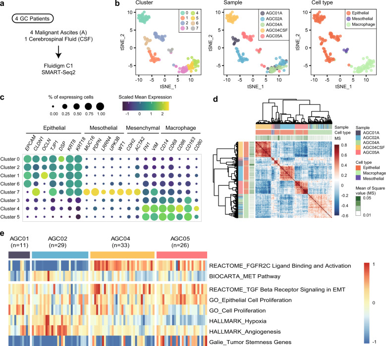
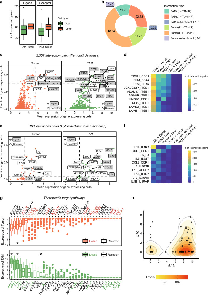
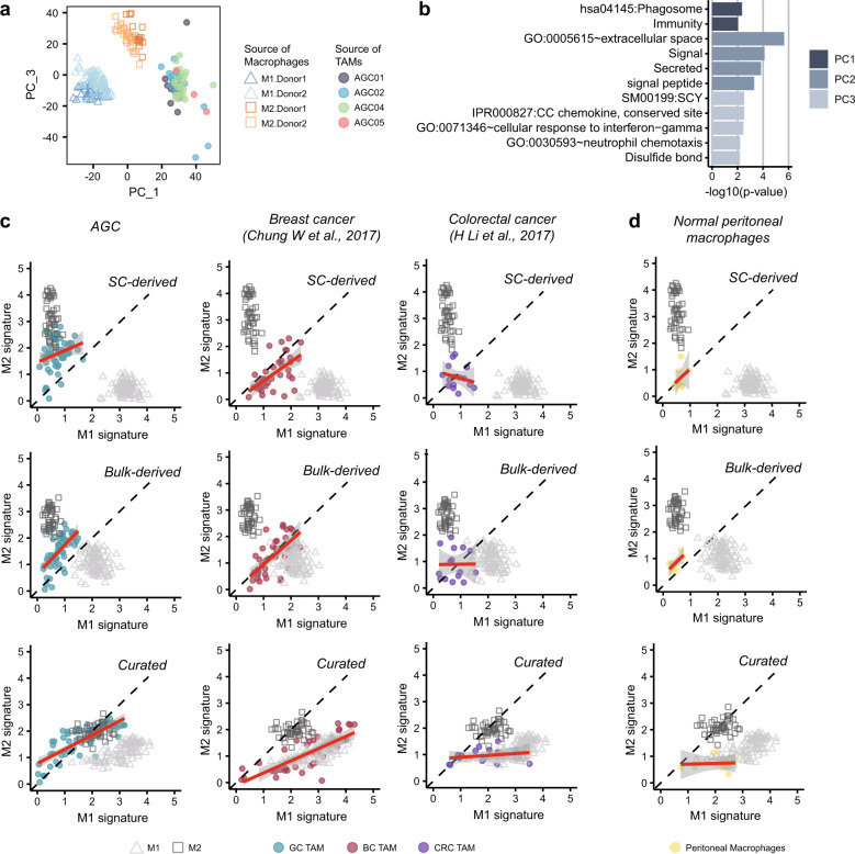
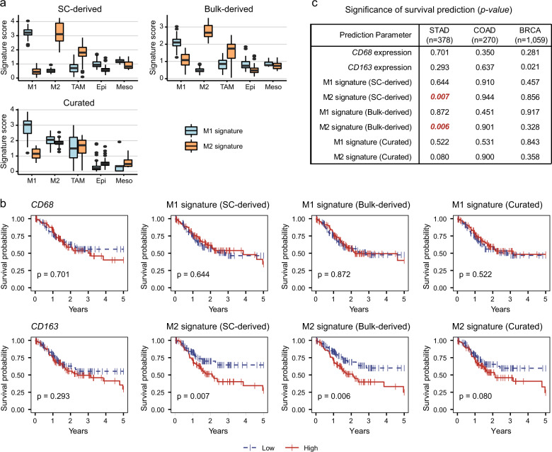
Gastric cancer (GC) patients develop malignant ascites as the disease progresses owing to peritoneal metastasis. GC patients with malignant ascites have a rapidly deteriorating clinical course with short survival following the onset of malignant ascites. Better optimized treatment strategies for this subset of patients are needed. To define the cellular characteristics of malignant ascites of GC, we used single-cell RNA sequencing to characterize tumor cells and tumor-associated macrophages (TAMs) from four samples of malignant ascites and one sample of cerebrospinal fluid. Reference transcriptomes for M1 and M2 macrophages were generated by in vitro differentiation of healthy blood-derived monocytes and applied to assess the inflammatory properties of TAMs. We analyzed 180 cells, including tumor cells, macrophages, and mesothelial cells. Dynamic exchange of tumor-promoting signals, including the CCL3-CCR1 or IL1B-IL1R2 interactions, suggests macrophage recruitment and anti-inflammatory tuning by tumor cells. By comparing these data with reference transcriptomes for M1-type and M2-type macrophages, we found noninflammatory characteristics in macrophages recovered from the malignant ascites of GC. Using public datasets, we demonstrated that the single-cell transcriptome-driven M2-specific signature was associated with poor prognosis in GC. Our data indicate that the anti-inflammatory characteristics of TAMs are controlled by tumor cells and present implications for treatment strategies for GC patients in which combination treatment targeting cancer cells and macrophages may have a reciprocal synergistic effect.


Conflict of interest statement

The authors declare that they have no conflict of interest.

Figures

Fig. 1
Fig. 1. Characterization of tumor cells and tumor-associated macrophages in malignant ascites.
a Scheme of data generation in the study. b Dimension reduction using t-distributed stochastic neighbor embedding (tSNE) separates noncancerous cell clusters and patient-specific cancer cell clusters. Each dot represents a cell, and each cell is colored by its SNN cluster (left), sample origin (middle), or cell type (right). c The expression of marker genes for epithelial cells, peritoneal mesothelial cells, mesenchymal cells, and macrophages clarified the cell type of the clusters. d Pearson’s correlation coefficient matrix of the CEP values. e Heatmap of tumor characteristics in the tumor cells. Each column represents a single cell.
Fig. 2
Fig. 2. Tumor-promoting interactions of cancer cells and macrophages in malignant ascites.
a The number of expressed ligand (left) or receptor (right) genes from the FANTOM5 database in each cell. b Putative intercellular interactions between two other cells are much more prominent than was putative self-sufficient signaling. Intercellular interaction types were indicated by ‘ligand-expressing cell (L)>receptor-expressing cell (R)’. c Strongly and commonly expressed interacting genes in tumors (left) or TAMs (right). The genes with average expression over quantile 0.95 and a fraction of expressing cells over 0.75 were labeled. d Top 10 abundant interaction pairs (>10,224 pairs in each interaction) related to c. e Strongly and commonly expressed interacting genes among the chemokine-related interactions (Supplementary Table 1a). Genes with average expression over quantile 0.5 and a fraction of expressing cells over 0.1 were labeled. f Top 10 abundant interaction pairs (>406 pairs in each interaction) related to e. g Expression level of the genes associated with macrophage function (Supplementary Table 1b) indicates high expression of CCL3, IL1B, and CCR1 in macrophages and IL1R2 in tumor cells (marked with a star). h Coexpression of IL1B and IL10 in TAMs. The x and y-axes show the expression levels of IL1B and IL10, respectively. Each dot represents a cell, and each cell was colored based on the sample origin. Expressed genes were evaluated based on threshold 1 in a–f.
Fig. 3
Fig. 3. M1- and M2-signature genes at single-cell resolution.
a Scheme of the in vitro differentiation of M1-type and M2-type macrophages. b Morphological changes during differentiation. c FACS analysis of the M1-specific marker CD80 and the M2-specific marker CD163 in differentiated M1 and M2 cells. The examined samples are colored with borders in a (black for monocytes, blue for M1 macrophages, and orange for M2 macrophages). d Unsupervised PCA primarily separated M1 and M2 macrophages by the first principal component. Cells are colored by cluster (upper) or sample origin (lower). e Gene expression levels of CD80 and CD163 in single-cell RNA-seq data confirmed M1 and M2 polarization. The thick black line indicates the median value of each sample. f M1-specific or M2-specific signature genes were extracted by overlapping differentially expressed genes from both donors. g Comparison with published gene sets derived from bulk M1 and M2 signatures. Only 29 genes were repeatedly extracted from single-cell and bulk signatures.
Fig. 4
Fig. 4. In vitro differentiated M1 and M2 transcriptomes show the M1-like and M2-like features of TAMs.
a PCA grouped TAMs into a separate cluster from the reference M1 or M2 cells. Cells were colored based on the origin of the sample. b The GO terms describing each principal component show the functional difference between macrophage types (Bonferroni corrected p-value < 0.01). The top 50 genes on each principal component were analyzed by DAVID 6.8. c Two-dimensional dot plots for M1 and M2 signature evaluation were constructed for three cancer types using three different gene sets: single-cell transcriptome-derived signatures (top), bulk transcriptome-derived signatures (middle), and curated signatures (bottom). The x-axis indicates expression in the M1 signature, and the y-axis indicates expression in the M2 signature. The red line represents the simple linear regression line, and the gray area represents the confidence interval. d The M1–M2 signature 2-D plot estimates the M1-like and M2-like phenotypes of normal peritoneal macrophages.
Fig. 5
Fig. 5. The M2 phenotype of TAMs can be applied to predict the prognosis in gastric cancer.
a Transcriptome-driven M2 signatures, not curated signatures, are high exclusively in M2-type cells. M1 reference M1 cells, M2 reference M2 cells, TAM macrophages from GC, Epi epithelial cells from GC, Meso mesothelial cells from GC. b Overall survival prediction was performed using the TCGA STAD dataset (n = 378) and either signature gene sets or canonical macrophage markers, such as CD68 and CD163. The ‘high’ group (red) and ‘low’ group (blue) were determined by the 25th and 75th percentiles, respectively. p indicates the p-value of log-rank test. c Summary table of p-values for overall survival using the TCGA dataset for the three cancer types (STAD, n = 378; COAD, n = 270; and BRCA, n = 1059).

References

    1. Cancer Genome Atlas Research, N. Comprehensive molecular characterization of gastric adenocarcinoma. Nature. 2014;513:202–209. - PMC - PubMed
    1. Cristescu R, et al. Molecular analysis of gastric cancer identifies subtypes associated with distinct clinical outcomes. Nat. Med. 2015;21:449–456. - PubMed
    1. Chau I, et al. Multivariate prognostic factor analysis in locally advanced and metastatic esophago-gastric cancer-pooled analysis from three multicenter, randomized, controlled trials using individual patient data. J. Clin. Oncol. 2004;22:2395–2403. - PubMed
    1. Kim ST, et al. Comprehensive molecular characterization of clinical responses to PD-1 inhibition in metastatic gastric cancer. Nat. Med. 2018;24:1449–1458. - PubMed
    1. Lee J, et al. Development of mesenchymal subtype gene signature for clinical application in gastric cancer. Oncotarget. 2017;8:66305–66315. - PMC - PubMed

Publication types

MeSH terms