

Quantification of myocardial strain assessed by cardiovascular magnetic resonance feature tracking in healthy subjects-influence of segmentation and analysis software
- PMID: 33277669
- PMCID: PMC8128822
- DOI: 10.1007/s00330-020-07539-5
Quantification of myocardial strain assessed by cardiovascular magnetic resonance feature tracking in healthy subjects-influence of segmentation and analysis software
Abstract
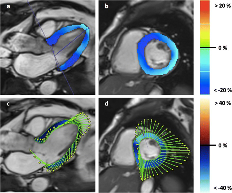
Objectives: Quantification of myocardial deformation by feature tracking is of growing interest in cardiovascular magnetic resonance. It allows the assessment of regional myocardial function based on cine images. However, image acquisition, post-processing, and interpretation are not standardized. We aimed to assess the influence of segmentation procedure such as slice selection and different types of analysis software on values and quantification of myocardial strain in healthy adults.
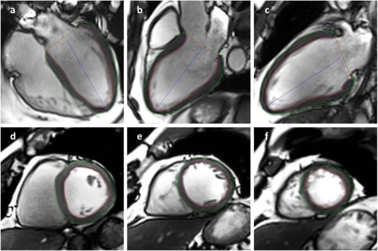
Methods: Healthy volunteers were retrospectively analyzed. Post-processing was performed using CVI42 and TomTec. Longitudinal and radialLong axis (LAX) strain were quantified using 4-chamber-view, 3-chamber-view, and 2-chamber-view. Circumferential and radialShort axis (SAX) strain were assessed in basal, midventricular, and apical short-axis views and using full coverage. Global and segmental strain values were compared to each other regarding their post-processing approach and analysis software package.
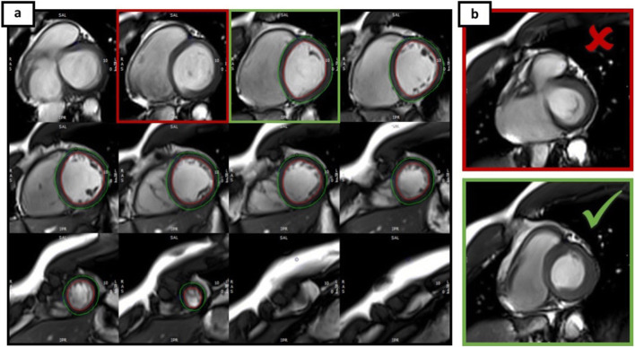
Results: We screened healthy volunteers studied at 1.5 or 3.0 T and included 67 (age 44.3 ± 16.3 years, 31 females). Circumferential and radialSAX strain values were different between a full coverage approach vs. three short slices (- 17.6 ± 1.8% vs. - 19.2 ± 2.3% and 29.1 ± 4.8% vs. 34.6 ± 7.1%). Different analysis software calculated significantly different strain values. Within the same vendor, different field strengths (- 17.0 ± 2.1% at 1.5 T vs. - 17.0 ± 1.7% at 3 T, p = 0.845) did not influence the calculated global longitudinal strain (GLS), and were similar in gender (- 17.4 ± 2.0% in females vs. - 16.6 ± 1.8% in males, p = 0.098). Circumferential and radial strain were different in females and males (circumferential strain - 18.2 ± 1.7% vs. - 17.1 ± 1.8%, p = 0.029 and radial strain 30.7 ± 4.7% vs. 27.8 ± 4.6%, p = 0.047).
Conclusions: Myocardial deformation assessed by feature tracking depends on segmentation procedure and type of analysis software. CircumferentialSAX and radialSAX depend on the number of slices used for feature tracking analysis. As known from other imaging modalities, GLS seems to be the most stable parameter. During follow-up studies, standardized conditions should be warranted. Trial registration Retrospectively registered KEY POINTS: • Myocardial deformation assessed by feature tracking depends on the segmentation procedure. • Global myocardial strain values differ significantly among vendors. • Standardization in post-processing using CMR feature tracking is essential.
Keywords: Healthy volunteers; Left ventricular function; Magnetic resonance imaging; Myocard; Software.
Conflict of interest statement
The authors of this manuscript declare no relationships with any companies whose products or services may be related to the subject matter of the article.
Figures


Similar articles
-
Age- and gender-related normal left ventricular deformation assessed by cardiovascular magnetic resonance feature tracking.J Cardiovasc Magn Reson. 2015 Mar 10;17(1):25. doi: 10.1186/s12968-015-0123-3. J Cardiovasc Magn Reson. 2015. PMID: 25890093 Free PMC article.
-
Global and regional left ventricular myocardial deformation measures by magnetic resonance feature tracking in healthy volunteers: comparison with tagging and relevance of gender.J Cardiovasc Magn Reson. 2013 Jan 18;15(1):8. doi: 10.1186/1532-429X-15-8. J Cardiovasc Magn Reson. 2013. PMID: 23331550 Free PMC article.
-
The intra-observer reproducibility of cardiovascular magnetic resonance myocardial feature tracking strain assessment is independent of field strength.Eur J Radiol. 2013 Feb;82(2):296-301. doi: 10.1016/j.ejrad.2012.11.012. Epub 2012 Dec 12. Eur J Radiol. 2013. PMID: 23246014
-
Myocardial Strain Measurements Derived From MR Feature-Tracking: Influence of Sex, Age, Field Strength, and Vendor.JACC Cardiovasc Imaging. 2024 Apr;17(4):364-379. doi: 10.1016/j.jcmg.2023.05.019. Epub 2023 Jul 19. JACC Cardiovasc Imaging. 2024. PMID: 37480906
-
Myocardial deformation assessment using cardiovascular magnetic resonance-feature tracking technique.Br J Radiol. 2017 Dec;90(1080):20170072. doi: 10.1259/bjr.20170072. Epub 2017 Oct 9. Br J Radiol. 2017. PMID: 28830199 Free PMC article. Review.
Cited by
-
Different Impacts on the Heart After COVID-19 Infection and Vaccination: Insights From Cardiovascular Magnetic Resonance.Front Cardiovasc Med. 2022 Jul 14;9:916922. doi: 10.3389/fcvm.2022.916922. eCollection 2022. Front Cardiovasc Med. 2022. PMID: 35911510 Free PMC article.
-
Right ventricular function declines prior to left ventricular ejection fraction in hypertrophic cardiomyopathy.J Cardiovasc Magn Reson. 2022 Jun 13;24(1):36. doi: 10.1186/s12968-022-00868-y. J Cardiovasc Magn Reson. 2022. PMID: 35692049 Free PMC article.
-
Layer-specific fast strain-encoded cardiac magnetic resonance imaging aids in the identification and discrimination of acute myocardial injury: a prospective proof-of-concept study.J Cardiovasc Magn Reson. 2024 Summer;26(1):101001. doi: 10.1016/j.jocmr.2024.101001. Epub 2024 Jan 19. J Cardiovasc Magn Reson. 2024. PMID: 38244931 Free PMC article.
-
Impact of myocardial deformation on risk prediction in patients following acute myocardial infarction.Front Cardiovasc Med. 2023 Aug 10;10:1199936. doi: 10.3389/fcvm.2023.1199936. eCollection 2023. Front Cardiovasc Med. 2023. PMID: 37636296 Free PMC article.
-
Cardiac Magnetic Resonance Relaxometry Parameters, Late Gadolinium Enhancement, and Feature-Tracking Myocardial Longitudinal Strain in Patients Recovered from COVID-19.J Cardiovasc Dev Dis. 2023 Jun 29;10(7):278. doi: 10.3390/jcdd10070278. J Cardiovasc Dev Dis. 2023. PMID: 37504534 Free PMC article.
References
-
- Barreiro-Perez M, Curione D, Symons R, Claus P, Voigt JU, Bogaert J. Left ventricular global myocardial strain assessment comparing the reproducibility of four commercially available CMR-feature tracking algorithms. Eur Radiol. 2018;28:5137–5147. - PubMed
-
- Plana JC, Galderisi M, Barac A, et al. Expert consensus for multimodality imaging evaluation of adult patients during and after cancer therapy: a report from the American Society of Echocardiography and the European Association of Cardiovascular Imaging. Eur Heart J Cardiovasc Imaging. 2014;15:1063–1093. - PMC - PubMed
-
- Celutkiene J, Plymen CM, Flachskampf FA, et al. Innovative imaging methods in heart failure: a shifting paradigm in cardiac assessment. Position statement on behalf of the Heart Failure Association of the European Society of Cardiology. Eur J Heart Fail. 2018;20:1615–1633. - PubMed
-
- Schuster A, Paul M, Bettencourt N, et al. Cardiovascular magnetic resonance myocardial feature tracking for quantitative viability assessment in ischemic cardiomyopathy. Int J Cardiol. 2013;166:413–420. - PubMed
-
- Buss SJ, Emami M, Mereles D, et al. Longitudinal left ventricular function for prediction of survival in systemic light-chain amyloidosis: incremental value compared with clinical and biochemical markers. J Am Coll Cardiol. 2012;60:1067–1076. - PubMed
MeSH terms
LinkOut - more resources
Full Text Sources
Research Materials
Miscellaneous
