

Decellularized tissue-engineered heart valves calcification: what do animal and clinical studies tell us?
- PMID: 33278023
- PMCID: PMC7719105
- DOI: 10.1007/s10856-020-06462-x
Decellularized tissue-engineered heart valves calcification: what do animal and clinical studies tell us?
Abstract
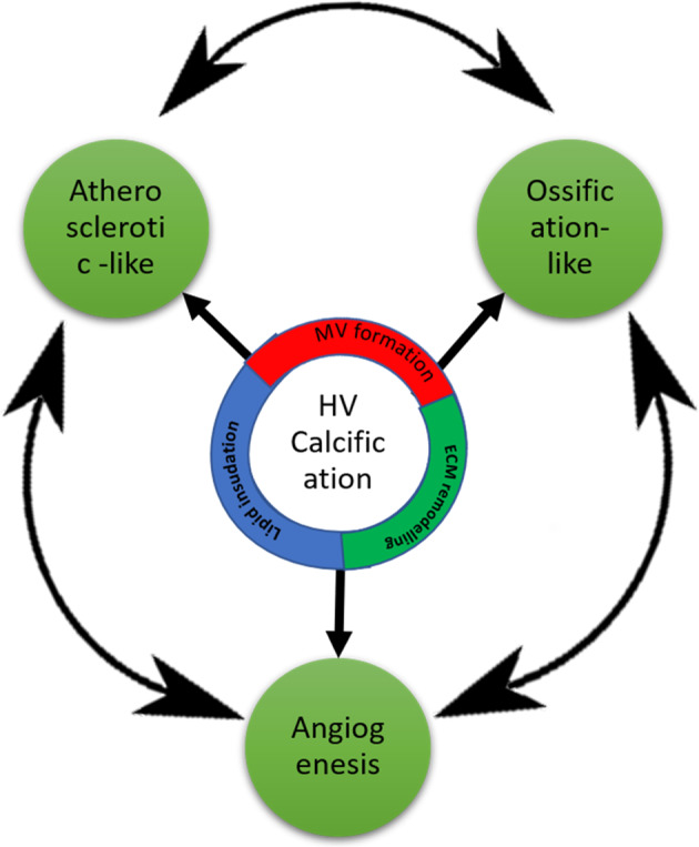
Cardiovascular diseases are the first cause of death worldwide. Among different heart malfunctions, heart valve failure due to calcification is still a challenging problem. While drug-dependent treatment for the early stage calcification could slow down its progression, heart valve replacement is inevitable in the late stages. Currently, heart valve replacements involve mainly two types of substitutes: mechanical and biological heart valves. Despite their significant advantages in restoring the cardiac function, both types of valves suffered from serious drawbacks in the long term. On the one hand, the mechanical one showed non-physiological hemodynamics and the need for the chronic anticoagulation therapy. On the other hand, the biological one showed stenosis and/or regurgitation due to calcification. Nowadays, new promising heart valve substitutes have emerged, known as decellularized tissue-engineered heart valves (dTEHV). Decellularized tissues of different types have been widely tested in bioprosthetic and tissue-engineered valves because of their superior biomechanics, biocompatibility, and biomimetic material composition. Such advantages allow successful cell attachment, growth and function leading finally to a living regenerative valvular tissue in vivo. Yet, there are no comprehensive studies that are covering the performance of dTEHV scaffolds in terms of their efficiency for the calcification problem. In this review article, we sought to answer the question of whether decellularized heart valves calcify or not. Also, which factors make them calcify and which ones lower and/or prevent their calcification. In addition, the review discussed the possible mechanisms for dTEHV calcification in comparison to the calcification in the native and bioprosthetic heart valves. For this purpose, we did a retrospective study for all the published work of decellularized heart valves. Only animal and clinical studies were included in this review. Those animal and clinical studies were further subcategorized into 4 categories for each depending on the effect of decellularization on calcification. Due to the complex nature of calcification in heart valves, other in vitro and in silico studies were not included. Finally, we compared the different results and summed up all the solid findings of whether decellularized heart valves calcify or not. Based on our review, the selection of the proper heart valve tissue sources (no immunological provoking residues), decellularization technique (no damaged exposed residues of the decellularized tissues, no remnants of dead cells, no remnants of decellularizing agents) and implantation techniques (avoiding suturing during the surgical implantation) could provide a perfect anticalcification potential even without in vitro cell seeding or additional scaffold treatment.
Conflict of interest statement
The authors declare that they have no conflict of interest.
Figures
References
-
- Otto CM, Prendergast, B. Aortic-valve stenosis - From patients at risk to severe valve obstruction. N Engl J Med. 2014. 10.1056/NEJMra1313875 - PubMed
-
- Rajamannan NM, Evans FJ, Aikawa E, Grande-Allen KJ, Demer LL, Heistad DD, et al. Calcific aortic valve disease: Not simply a degenerative process: a review and agenda for research from the national heart and lung and blood institute aortic stenosis working group. Circulation. 2011. 10.1161/CIRCULATIONAHA.110.006767 - PMC - PubMed
-
- Steinberg DH. Aortic valve calcification: moving toward the root of the problem. J Am Coll Cardiol. 2019. 10.1016/j.jacc.2018.11.016 - PubMed
-
- Mavrilas D, Apostolakis, E, Koutsoukos P. Prosthetic aortic valves: a surgical and bioengineering approach. In Aortic valve surgery; 2011. 10.5772/20860
Publication types
MeSH terms
Substances
LinkOut - more resources
Full Text Sources
