. 2021 Feb 3;109(3):461-472.e5.
doi: 10.1016/j.neuron.2020.11.010. Epub 2020 Dec 4.

Area Postrema Cell Types that Mediate Nausea-Associated Behaviors

Affiliations

Area Postrema Cell Types that Mediate Nausea-Associated Behaviors

Chuchu Zhang et al. Neuron. .

Abstract

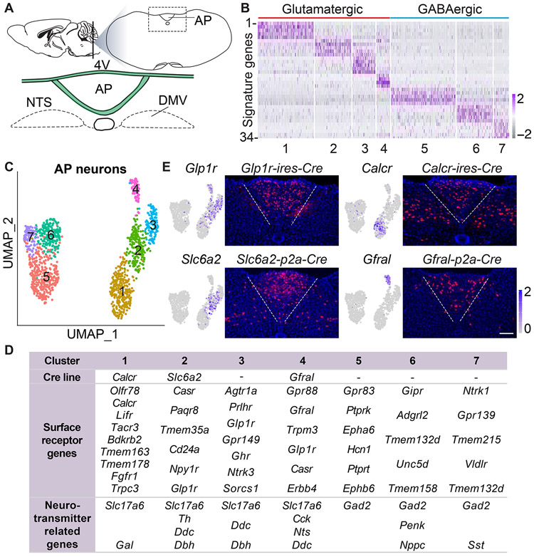
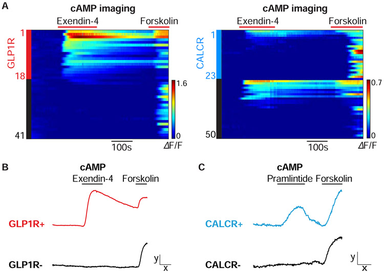
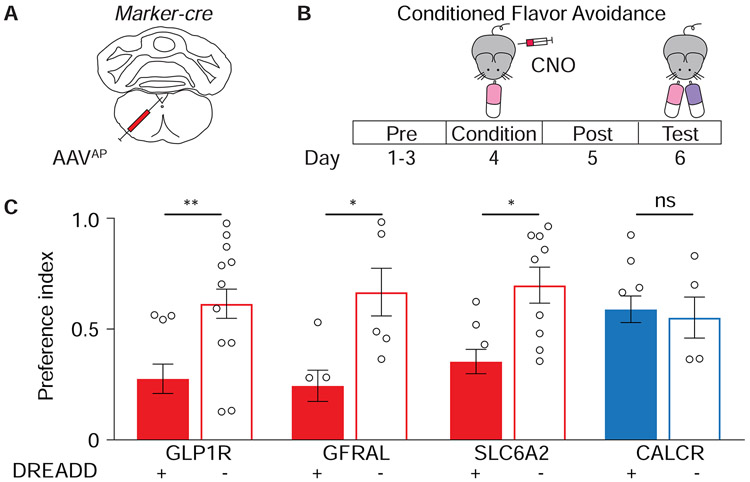
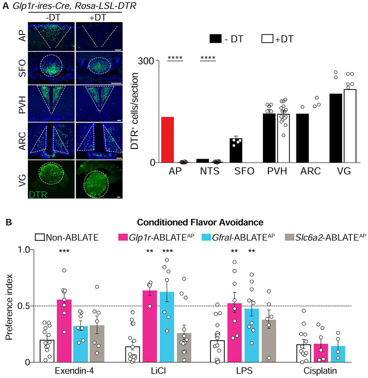
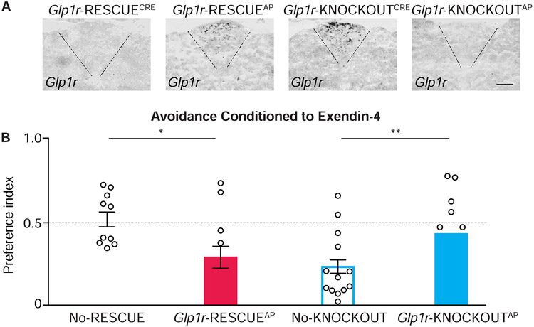
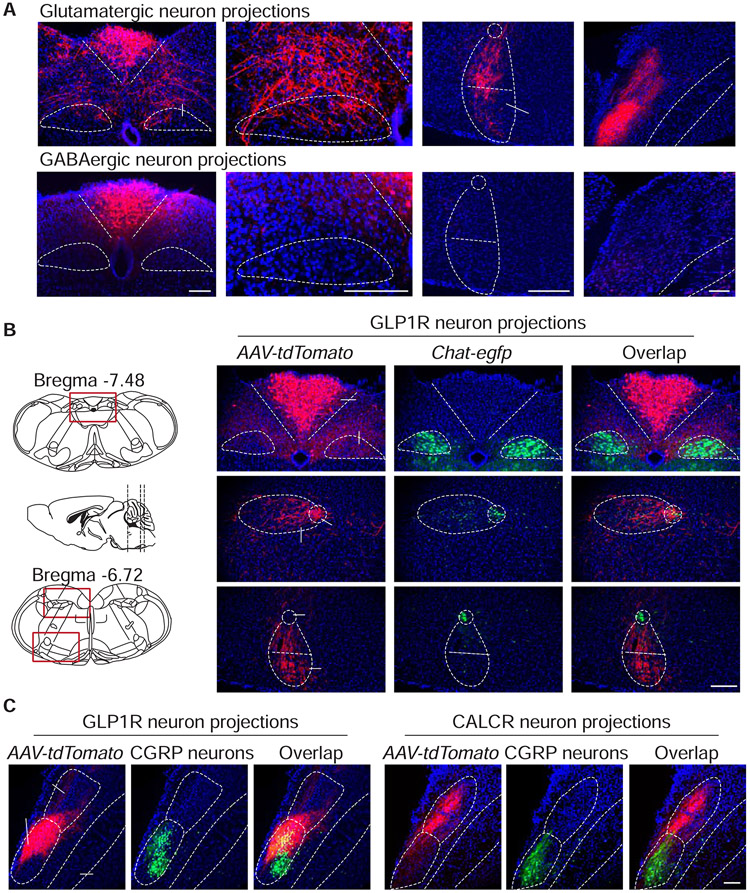
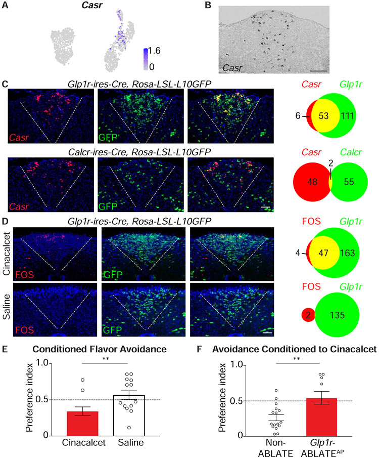
Nausea, the unpleasant sensation of visceral malaise, remains a mysterious process. The area postrema is implicated in some nausea responses and is anatomically privileged to detect blood-borne signals. To investigate nausea mechanisms, we built an area postrema cell atlas through single-nucleus RNA sequencing, revealing a few neuron types. Using mouse genetic tools for cell-specific manipulation, we discovered excitatory neurons that induce nausea-related behaviors, with one neuron type mediating aversion imposed by multiple poisons. Nausea-associated responses to agonists of identified area postrema receptors were observed and suppressed by targeted cell ablation and/or gene knockout. Anatomical mapping revealed a distributed network of long-range excitatory but not inhibitory projections with subtype-specific patterning. These studies reveal the basic organization of area postrema nausea circuitry and provide a framework toward understanding and therapeutically controlling nausea.

Keywords: Calcium sensing receptor (CaSR); GDF15; GFRAL; Glucagon-like peptide 1 receptor (GLP1R); circumventricular organs; conditioned flavor avoidance; emesis; exendin-4; lithlum chloride; parabrachial nucleus CGRP neurons.


Conflict of interest statement

Declaration of Interests S.D.L. is a consultant for Kallyope, Inc.

Figures

Figure 1.
Figure 1.. Genetic control over an area postrema cell atlas
(A) Cartoon indicating area postrema (AP) location in the brainstem; 4V: fourth ventricle; AP: area postrema; DMV: dorsal motor nucleus of the vagus. (B) Normalized expression levels of 34 cluster-defining signature genes (see Table S1 for gene names) across all cells from clusters 1-7. (C) Uniform manifold approximation and projection (UMAP) plots based on single-cell transcriptome data showing seven area postrema neuron types (color coded) including excitatory (clusters 1-4) and inhibitory neurons (clusters 5-7). (D) Receptor and neurotransmitter-related genes expressed in area postrema neuron types. (E) UMAP plots indicating expression of Cre-targeted genes in area postrema neurons (left). Cre lines indicated were crossed to a Rosa26-LSL-tdTomato reporter, and native fluorescence visualized in coronal area postrema cryosections (right), scale bars: 100 μm. See also Figure S1, S2 and Table S1.
Figure 2.
Figure 2.. Imaging responses of area postrema neurons
(A) Area postrema neurons of Glp1r-ires-Cre; Rosa26-LSL-tdTomato (left) or Calcr-ires-Cre; Rosa26-LSL-tdTomato (right) mice were acutely dissociated and transfected with the cAMP sensor cADDis. Rows indicate responses (ΔF/F, color scale) of individual neurons over time, with exendin-4 (100 nM) and forskolin (25 μm) applied at times indicated (red bars). Y-axis colored bars (left: red; right: blue) and black bars indicated tdTomato-positive and tdTomato-negative neurons respectively. (B, C) Area postrema neurons were harvested from Glp1r-ires-Cre; Rosa26-LSL-tdTomato (B) or Calcr-ires-Cre; Rosa26-LSL-tdTomato mice (C) for transfection-based cAMP imaging. Representative responses of GLP1R-positive (red), CALCR-positive (blue), and reporter-negative (black) neurons are shown to exendin-4 (100 nM), pramlintide (100 nM), and forskolin (25 μM) x: 100 seconds, y: 0.5 ΔF/F.
Figure 3.
Figure 3.. Multiple area postrema neuron types promote flavor avoidance
(A) Cartoon of AAV infection in area postrema. (B) Timeline of behavioral assay for conditioned flavor avoidance. (C) Glp1r-ires-Cre (GLP1R), Gfral-p2a-Cre (GFRAL), Slc6a2-p2a-Cre (SLC6A2), and Calcr-ires-Cre (CALCR) mice were injected with AAVs encoding Cre-dependent Gαs-DREADD-mCherry (+) or tdTomato (−), and analyzed for CNO-evoked flavor avoidance. (n=5-15, mean ± sem, circles: individual data points, *p<.05, **p<.01. See also Figure S3, S4.
Figure 4.
Figure 4.. Neuron ablation eliminates behavioral responses to visceral poisons
(A) The area postrema of Glp1r-ires-Cre; Rosa26-LSL-DTR mice was injected with DT (+DT) or saline (−DT). Immunohistochemistry for DTR (left) and quantification of DTR-expressing cells (right) in coronal brain cryosections containing area postrema (AP), subfornical organ (SFO), paraventricular hypothalamus (PVH), and arcuate nucleus (ARC) or in wholemounts of vagal ganglia (VG), scale bar: 100 μm, n=4-21 sections from 4 mice (−DT) or 14 mice (+DT), mean ± sem, circles: individual data points, ****p<.0001. (B) The ability of exendin-4, lithium chloride (LiCl), lipopolysaccharide (LPS), and cisplatin to condition flavor avoidance in Non-ABLATE, Glp1r-ABLATEAP, Slc6a2-ABLATEAP, and Gfral-ABLATEAP mice, n=4-18, mean ± sem, circles: individual data points, **p<.01, ***p<.001, statistical comparisons to non-ABLATE mice. See also Figure S5.
Figure 5.
Figure 5.. Cell-specific knockout and rescue of Glp1r impacts flavor avoidance
(A) In situ hybridization to detect Glp1r mRNA in coronal area postrema cryosections from mice indicated, scale bar: 100 μm. (B) The ability of exendin-4 to condition flavor avoidance was measured in mice indicated, n=11-18, mean ± sem, circles: individual data points, *p<.05, **p<.01. See also Figure S5.
Figure 6.
Figure 6.. Cell-type specific projections of area postrema neurons
(A) AAV-Flex-tdTomato was injected into the area postrema of Slc17a6-ires-Cre (top) and Gad2-ires-Cre (bottom) mice, and tdTomato-positive fibers were visualized by immunohistochemistry. (B) AAV-Flex-tdTomato was injected into the area postrema of Glp1r-ires-Cre; Chat-gfp mice. GFP was visualized by native fluorescence and tdTomato-positive fibers were visualized by immunohistochemistry. (C) AAV-Flex-tdTomato was injected into the area postrema of Glp1r-ires-Cre; Calca-gfp and Calcr-ires-Cre; Calca-gfp mice. GFP was visualized by native fluorescence (CGRP neurons) and tdTomato-positive fibers were visualized by immunohistochemistry, AP: area postrema; DMV: dorsal motor nucleus of the vagus; Amb: nucleus ambiguus; RVLM: rostral ventrolateral medulla; PBN: parabrachial nucleus; dl: dorsolateral; el: external lateral; scp: superior cerebellar peduncle; scale bars: 100 μm. See also Figure S6, S7.
Figure 7.
Figure 7.. A neural basis for behavioral aversion conditioned by cinacalcet
(A) UMAP plot depicting Casr expression across area postrema neurons. (B) In situ hybridization to detect Casr mRNA in coronal area postrema cryosections, scale bar: 100 μm. (C) Two-color expression analysis showing in situ hybridization to detect Casr mRNA (red) and native GFP fluorescence (green) in coronal area postrema cryosections of Glp1r-ires-Cre; Rosa26-LSL-L10GFP and Calcr-ires-Cre; Rosa26-LSL-L10GFP mice, scale bar: 100 μm. The numbers of co-labeled (yellow) or individually labeled (red, green) cells were counted (right). (D) Fos immunohistochemistry (red) and native GFP fluorescence (green) were analyzed in coronal area postrema cryosections after intraperitoneal injection of cinacalcet or saline alone in Glp1r-ires-Cre; Rosa26-LSL-L10GFP mice, scale bar: 100 μm. (E) The ability of intraperitoneal cinacalcet or saline to condition flavor avoidance was measured in wild-type mice, n=11-14, mean ± sem, circles: individual data points, **p<.01. (F) The ability of cinacalcet to condition flavor avoidance was measured in Non-ABLATE and Glp1r-ABLATEAP mice, n=10-16, mean ± sem, circles: individual data points, **p<.01.

Comment in

References

    1. Adams JM, Pei H, Sandoval DA, Seeley RJ, Chang RB, Liberles SD, and Olson DP (2018). Liraglutide Modulates Appetite and Body Weight Through Glucagon-Like Peptide 1 Receptor-Expressing Glutamatergic Neurons. Diabetes 67, 1538–1548. - PMC - PubMed
    1. Andrews PL (1992). Physiology of nausea and vomiting. Br J Anaesth 69, 2S–19S. - PubMed
    1. Baggio LL, and Drucker DJ (2007). Biology of incretins: GLP-1 and GIP. Gastroenterology 132, 2131–2157. - PubMed
    1. Barth SW, Riediger T, Lutz TA, and Rechkemmer G (2004). Peripheral amylin activates circumventricular organs expressing calcitonin receptor a/b subtypes and receptor-activity modifying proteins in the rat. Brain research 997, 97–102. - PubMed
    1. Borison HL (1989). Area postrema: chemoreceptor circumventricular organ of the medulla oblongata. Progress in neurobiology 32, 351–390. - PubMed

Publication types

MeSH terms

Substances