. 2021 May;18(5):801-810.
doi: 10.1016/j.hrthm.2020.11.026. Epub 2020 Dec 3.

A circadian clock in the sinus node mediates day-night rhythms in Hcn4 and heart rate

Affiliations

A circadian clock in the sinus node mediates day-night rhythms in Hcn4 and heart rate

Alicia D'Souza et al. Heart Rhythm. 2021 May.

Abstract

Background: Heart rate follows a diurnal variation, and slow heart rhythms occur primarily at night.

Objective: The lower heart rate during sleep is assumed to be neural in origin, but here we tested whether a day-night difference in intrinsic pacemaking is involved.

Methods: In vivo and in vitro electrocardiographic recordings, vagotomy, transgenics, quantitative polymerase chain reaction, Western blotting, immunohistochemistry, patch clamp, reporter bioluminescence recordings, and chromatin immunoprecipitation were used.

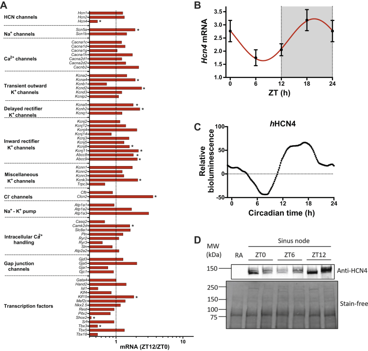
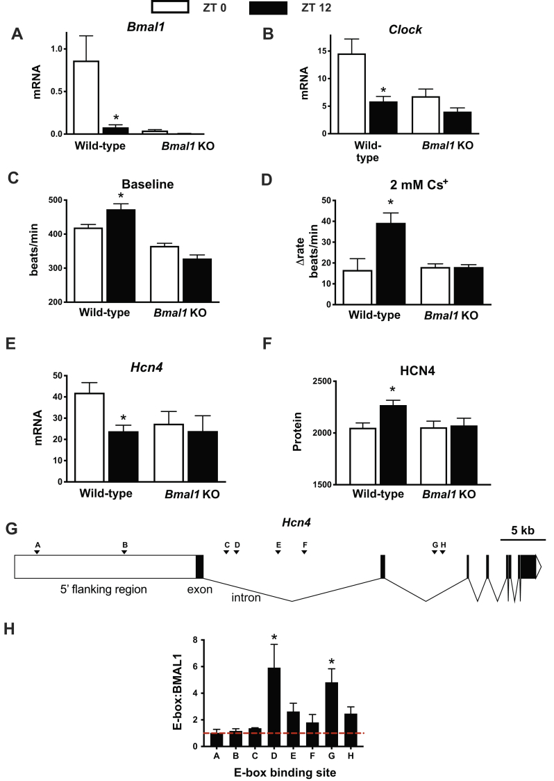
Results: The day-night difference in the average heart rate of mice was independent of fluctuations in average locomotor activity and persisted under pharmacological, surgical, and transgenic interruption of autonomic input to the heart. Spontaneous beating rate of isolated (ie, denervated) sinus node (SN) preparations exhibited a day-night rhythm concomitant with rhythmic messenger RNA expression of ion channels including hyperpolarization-activated cyclic nucleotide-gated potassium channel 4 (HCN4). In vitro studies demonstrated 24-hour rhythms in the human HCN4 promoter and the corresponding funny current. The day-night heart rate difference in mice was abolished by HCN block, both in vivo and in the isolated SN. Rhythmic expression of canonical circadian clock transcription factors, for example, Brain and muscle ARNT-Like 1 (BMAL1) and Cryptochrome (CRY) was identified in the SN and disruption of the local clock (by cardiomyocyte-specific knockout of Bmal1) abolished the day-night difference in Hcn4 and intrinsic heart rate. Chromatin immunoprecipitation revealed specific BMAL1 binding sites on Hcn4, linking the local clock with intrinsic rate control.

Conclusion: The circadian variation in heart rate involves SN local clock-dependent Hcn4 rhythmicity. Data reveal a novel regulator of heart rate and mechanistic insight into bradycardia during sleep.

Keywords: Bradycardia; Circadian rhythm; Pacemaking; Sinus node; Vagus nerve.


Figures

Figure 1
Figure 1
The day-night difference in average heart rate is independent of average physical activity and autonomic tone. A: Heart rate, PR interval, QRS duration, uncorrected QJ interval, and physical activity (measured using telemetry) in conscious mice (n = 9) over ∼6 days. Light and dark shaded regions represent light and dark phases in this and all similar figures. Timing of 1-hour light pulses is shown. In this and all similar figures, data are fit with a standard sine wave (red). Values are presented as mean ± SEM in this and all other figures (except in the case of physical activity, for which mean ± SD values are presented). B:In vivo heart rate and physical activity measured by telemetry at the times shown during 24-hour darkness (subjective day and night is shown) with the exception of a 1-hour light pulse delivered toward the start of the day (left) or night (right). The dotted red lines highlight the heart rate and physical activity at the end of the day-time light pulse, and the red arrows highlight the heart rate at the end of the nighttime light pulse. From the same experiment as in panel A. C:In vivo heart rate measured from anesthetized mice at ZT 0 and ZT 12 (n = 9 mice per time point) before (control) and after autonomic block by intraperitoneal injection of 1 mg/kg of atropine and 1 mg/kg of propranolol. D: Spontaneous beating rate of the mouse SN isolated at ZT 0 (n =8) and ZT 12 (n = 6). E:In vivo heart rate at ZT 0 and ZT 12 measured by telemetry in vagotomized rats (n = 7) at baseline (presurgery) and at 1, 3, and 7 days postsurgery. F:In vivo heart rate over 24 hours measured in telemetrized wild-type control (n = 21) and Girk4−/− (n = 23 mice). ∗P < .05. HCN4 = hyperpolarization activated cyclic nucleotide gated potassium channel 4; SN = sinus node; ZT = zeitgeber time.
Figure 2
Figure 2
Day-night rhythms in SN ion channel profile and Hcn4. A: Relative expression of transcripts in the SN at ZT 12 (vs ZT 0; n = 7/9 mice). The vertical line corresponds to 1, that is, no change. Values <1 correspond to a decrease at ZT 12 and >1 an increase, ∗P<0.05. B: Expression of Hcn4 mRNA (normalized to the expression of Tbp and Ipo8) in the SN at 4 time points over 24 hours (n = 6 mice at each time point). C: Representative LumiCycle data (Actimetrics, Wilmette, Illinois) (in arbitrary units) showing human Hcn4 promoter activity over 24 hours. D: Representative HCN4 Western blot from the SN dissected at ZT 0, ZT 6, and ZT 12. A right atrial tissue sample (RA) is also shown as a negative control; as expected, there is no HCN4 expression. Corresponding stain-free total protein gel was used for quantification shown in the lower panel. HCN4 = hyperpolarization activated cyclic nucleotide gated potassium channel 4; mRNA = messenger RNA; MW = molecular weight; SN = sinus node; ZT = zeitgeber time.
Figure 3
Figure 3
Day-night rhythm in If. A: Families of recordings of If made from SN cells isolated at ZT 0, ZT 6, and ZT 12. B: Current-voltage relationships for If recorded from SN cells isolated at ZT 0 (n = 38 cells per 4 mice), ZT 6 (n = 38 cells per 3 mice), and ZT 12 (n = 27 cells per 5 mice). C: HCN4 protein expression from the Western blot at ZT 0 (n = 10), ZT 6 (n = 5), and ZT 12 (n = 10). Data pooled from 2 sets of independent experiments and protein expression are normalized to those at ZT 0. D: Density of If at −120 mV at ZT 0 (n = 38 cells per 4 mice), ZT 6 (n = 38 cells per 3 mice), and ZT 12 (n = 27 cells per 5 mice). E: Amplitude of If at −120 mV at ZT 0, ZT 6, and ZT 12 (same data as in panel D). F: Cell capacitance at ZT 0, ZT 6, and ZT 12 (same data as in panel D). ∗P < .05. HCN4 = hyperpolarization activated cyclic nucleotide gated potassium channel 4; If = funny current; SN = sinus node; ZT = zeitgeber time.
Figure 4
Figure 4
Effect of HCN4 block on the heart rate. A: Representative electrographic recordings from telemetrized mice obtained at ZT 0 and ZT 12 at baseline and after the application of 6 mg/kg of ivabradine. B:In vivo heart rate of wild-type mice measured at ZT 0 and ZT 12 before and after the administration of 6 mg/kg of ivabradine (n = 3 mice per time point). C: Representative extracellular potential recordings from the SN isolated at ZT 0 and ZT 12 at baseline and after the application of 2 mM Cs+. D: Intrinsic heart rate of the isolated SN at ZT 0 and ZT 12 before and after the application of 2 mM Cs+ (n = 8 mice at baseline and 6 mice for Cs+ at ZT 0; n = 6 mice at baseline and 7 mice for Cs+ at ZT 12). ∗P < .05. HCN4 = hyperpolarization activated cyclic nucleotide gated potassium channel 4; SN = sinus node; ZT = zeitgeber time.
Figure 5
Figure 5
An intrinsic circadian clock in the SN. A: Relative expression of transcripts encoding key circadian clock components in the SN at ZT 12 vs ZT 0. The vertical line corresponds to 1, that is, no change. Values <1 correspond to a decrease at ZT 12 and >1 an increase (n = 7/9 mice). B: Expression of Bmal1 and Clock mRNA in the mouse SN at 4 time points over 24 hours (n = 6 at ZT 0, n = 6 at ZT 6; n = 7 at ZT 12 and n = 8 at ZT 18). C: Representative Per1 activity (reported by luciferase bioluminescence) in the isolated SN from Per1::LUC mice with a Cry1+/+Cry2+/+ or Cry1−/−Cry2−/− background. ∗P < .05. mRNA = messenger RNA; SN = sinus node; ZT = zeitgeber time.
Figure 6
Figure 6
BMAL1 modulates intrinsic heart rate and HCN4. A and B: Expression of Bmal1 (panel A) and Clock (panel B) at ZT 0 and ZT 12 in the SN of wild-type and Bmal1 KO mice (n = 6 at ZT 0 and n = 7 at ZT 12 for wild-type mice; n = 5 at ZT 0 and n = 6 at ZT 12 for Bmal1 KO mice). C: SN beating rate at ZT 0 and ZT 12 from wild-type (n = 8 at ZT 0 and n = 6 at ZT 12) and Bmal1 KO (n = 3 per time point) mice. D: Change in SN beating rate at ZT 0 and ZT 12 from wild-type (n = 8 at ZT 0 and n = 6 at ZT 12) and Bmal1 KO (n = 3 per time point) mice on application of 2 mM Cs+. E and F: Expression of Hcn4 mRNA (panel E; n = 6 at ZT 0 and n = 7 at ZT 12 for wild-type mice; n =5 at ZT 0 and n = 6 at ZT 12 for Bmal1 KO mice) and HCN4 protein (panel F; determined by immunohistochemistry; in arbitrary units; n = 56 sections at ZT 0 and n = 42 sections at ZT 12 for wild-type mice; n = 38 sections at ZT 0 and n = 46 sections at ZT 12 for Bmal1 KO mice; 3 mice per group) in the SN of mice at ZT 0 and ZT 12. G: Diagram of Hcn4 gene with 20 kb of the 5′ flanking region showing the position of 8 potential E-box binding sites (panels A–H). H: Eight potential E-box binding sites pulled down on immunoprecipitation of His-tagged BMAL1 from Cos-1 cells transfected with His-tagged Bmal1. Data are normalized to immunoprecipitation from untransfected control cells; the red dashed line equals 1 and is the baseline level. ∗P < .05 (binding site of interest vs binding site A; n = 2 replicate experiments). HCN4 = hyperpolarization activated cyclic nucleotide gated potassium channel 4; KO = knockout; mRNA = messenger RNA; SN = sinus node; ZT = zeitgeber time.

Comment in

  • Heartbeat music.
    Lakatta EG. Lakatta EG. Heart Rhythm. 2021 May;18(5):811-812. doi: 10.1016/j.hrthm.2021.01.011. Epub 2021 Jan 17. Heart Rhythm. 2021. PMID: 33465513 Free PMC article. No abstract available.

References

    1. Black N., D’Souza A., Wang Y. Circadian rhythm of cardiac electrophysiology, arrhythmogenesis, and the underlying mechanisms. Heart Rhythm. 2019;16:298–307. - PMC - PubMed
    1. Sutherland G.A. The pulse rate and range in health and disease during childhood. Q J Med. 1929;22:519–529.
    1. Vandewalle G., Middleton B., Rajaratnam S.M. Robust circadian rhythm in heart rate and its variability: influence of exogenous melatonin and photoperiod. J Sleep Res. 2007;16:148–155. - PubMed
    1. Monfredi O., Lyashkov A.E., Johnsen A.B. Biophysical characterisation of the under-appreciated and important relationship between heart rate variability and heart rate. Hypertension. 2014;64:1334–1343. - PMC - PubMed
    1. Boyett M., Wang Y., D’Souza A. CrossTalk opposing view: heart rate variability as a measure of cardiac autonomic responsiveness is fundamentally flawed. J Physiol. 2019;597:2599–2601. - PMC - PubMed

Publication types

MeSH terms

Substances