

Observations on the growth of temporalis muscle: A 3D CT imaging study
- PMID: 33280101
- PMCID: PMC8053578
- DOI: 10.1111/joa.13370
Observations on the growth of temporalis muscle: A 3D CT imaging study
Abstract
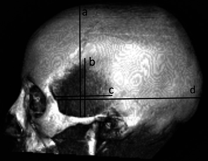
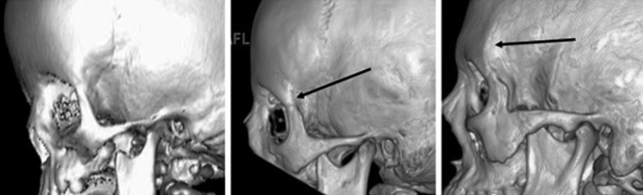
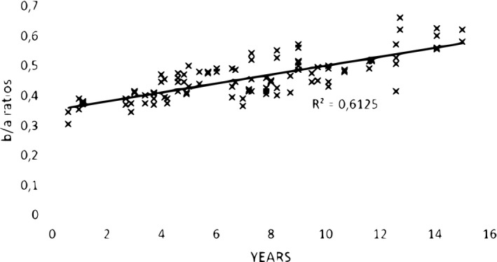
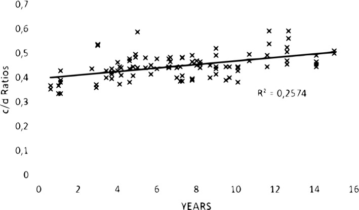
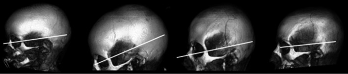
This study investigates the hypothesis that there is, during childhood, a disproportionate age-related expansion of the origin of temporalis muscle compared to the growth of the underlying skull. Lateral projections of 50 randomly selected 3D reformatted computerized tomographic (CT) scans (yielding 100 temporalis muscles) of children aged >0.6 to 15 years scanned for conditions that did not affect the shape of their head or face were windowed to provide the optimum delineation of temporalis muscle against the underlying bone. Vertical and anterior-posterior measurements of the muscle made independently by two observers were compared with those of the skull along the same planes. The development of the anterior temporal crest was also assessed. The intraclass correlation coefficient for differences in the measurements made by each observer ranged from good to excellent. The muscle and skull measurements were used to produce a ratio of muscle-to-skull lengths in both vertical and horizontal planes. Analysis of these ratios showed a statistically significant increase in the vertical reach of temporalis with age (Pearson correlation coefficient (R) =0.7826; p < 0.05) compared to the growth of the skull along the planes chosen for the study-but less so for its horizontal reach (R = 0.5073. p < .001). There were no significant differences between right/left or male/female measurements. There was also a substantial level of agreement between both observers in their assessment of the development of the temporal crest. The mean age of children in whom a fully formed temporal crest could be identified (10.6 years) was significantly greater (p < 0.001) than that of the 38 remaining subjects (6.0 years). These results confirm that there is, in response to increased masticatory/dietary demands during childhood, a disproportionate increase in the vertical and (to a lesser extent) horizontal reach of temporalis muscle over its origin from the temporal, frontal, sphenoid, and parietal bones compared the growth of the skull. It is proposed that surgical interference with this normal process is responsible for the soft tissue component of late-developing deformity that can occur following early (at 6-18 months of age) operations for the correction of trigonocephalic head shape associated with metopic synostosis.
Keywords: childhood; muscle growth; temporalis muscle.
© 2021 Anatomical Society.
Conflict of interest statement
The authors declare no conflicts of interest in the preparation of this paper.
Figures
References
-
- Chong, X. , Khoo, C.D. , Goh, H. , Rahman, F. & Shoji, Y. (2016) Effect of age on bite force. Journal of Oral Science, 58, 361–363. - PubMed
-
- Gisel, E.G. (1991) Effect of food texture on the development of chewing of children between six months and two years of age. Developmental Medicine and Child Neurology, 33, 69–79. - PubMed
-
- Joubert, D.M. (1955) Growth of muscle fibre in the fetal sheep. Nature, 175, 936–937. - PubMed
MeSH terms
LinkOut - more resources
Full Text Sources
