. 2021 Feb;27(2):49-58.
doi: 10.1089/ten.TEC.2020.0263. Epub 2021 Jan 20.

A Novel Three-Dimensional Skin Disease Model to Assess Macrophage Function in Diabetes

Affiliations

A Novel Three-Dimensional Skin Disease Model to Assess Macrophage Function in Diabetes

Avi Smith et al. Tissue Eng Part C Methods. 2021 Feb.

Abstract

A major challenge in the management of patients suffering from diabetes is the risk of developing nonhealing foot ulcers. Most in vitro methods to screen drugs for wound healing therapies rely on conventional 2D cell cultures that do not closely mimic the complexity of the diabetic wound environment. In addition, while three-dimensional (3D) skin tissue models of human skin exist, they have not previously been adapted to incorporate patient-derived macrophages to model inflammation from these wounds. In this study, we present a 3D human skin equivalent (HSE) model incorporating blood-derived monocytes and primary fibroblasts isolated from patients with diabetic foot ulcers (DFUs). We demonstrate that the monocytes differentiate into macrophages when incorporated into HSEs and secrete a cytokine profile indicative of the proinflammatory M1 phenotype seen in DFUs. We also show how the interaction between fibroblasts and macrophages in the HSE can guide macrophage polarization. Our findings take us a step closer to creating a human, 3D skin-like tissue model that can be applied to evaluate the response of candidate compounds needed for potential new foot ulcer therapies in a more complex tissue environment that contributes to diabetic wounds. Impact statement This study is the first to incorporate disease-specific, diabetic macrophages into a three-dimensional (3D) model of human skin. We show how to fabricate skin that incorporates macrophages with disease-specific fibroblasts to guide macrophage polarization. We also show that monocytes from diabetic patients can differentiate into macrophages directly in this skin disease model, and that they secrete a cytokine profile mimicking the proinflammatory M1 phenotype seen in diabetic foot ulcers. The data presented here indicate that this 3D skin disease model can be used to study macrophage-related inflammation in diabetes and as a drug testing tool to evaluate new treatments for the disease.

Keywords: diabetes; diabetic foot ulcer; human skin equivalent; macrophage; skin model.


Conflict of interest statement

The authors state no competing financial interests exist.

Figures

FIG. 1.
FIG. 1.
Development of an HSE tissue. (a) An acellular collagen layer is placed on the bottom of a 24 mm transwell membrane. (b) Fibroblasts with or without macrophages are mixed with bovine type 1 collagen and seeded on top. (c) The fibroblasts remodel and contract the collagen over the course of 1 week. (d) Neonatal human keratinocytes are seeded on top of the collagen matrix, where (e) they proliferate and begin differentiation. (f) The tissue is brought to an air-liquid interface to catalyze epithelial cornification. HSE, human skin equivalent. Color images are available online.
FIG. 2.
FIG. 2.
Monocytes isolated from Apheresis leukoreduction collars can be differentiated and polarized in 2D. (a) Monocytes seeded onto Petri dishes attached to the dishes and showed varied morphology after treatment with polarization factors. Scale bars: 200 μm. (b) Staining of macrophages indicated that all were positive for CD68, a pan-macrophage marker. HLADR staining was most present in macrophages treated with M1 polarization factors, CD163 was most present in macrophages treated with M2C polarization factors, and CD206 was most present in macrophages treated with M2A polarization factors. Scale bars: 100 μm. (c) The MFI by flow cytometry was highest for each population in the marker correlating with their polarization state. 2D, two-dimensional; MFI, mean fluorescence intensity. Color images are available online.
FIG. 3.
FIG. 3.
HSEs can be constructed with macrophages incorporated. H&E staining showed normal tissue development with a cellular dermal layer and full thickness epithelium with and without macrophages (a, b). Staining for K10 showed a differentiated epithelium both with and without macrophages (c, d). Scale bars: 100 μm. H&E, hematoxylin and eosin; K10, keratin 10. Color images are available online.
FIG. 4.
FIG. 4.
Macrophages persist in HSEs. (a) Immunofluorescent staining for the pan-macrophage marker CD11b (green), indicated that macrophages were present in HSEs at the end of development. Nonspecific CD68 staining was seen in the epithelium. Staining for the M1 marker HLADR, M2A marker CD206, and M2C marker CD163 (all red) showed positive cells in most tissues, regardless of macrophage polarization state before tissue formation. Tissues were counterstained with Dapi (blue). Yellow demonstrates overlap between green and red. (b) In HSEs where macrophages were incorporated unpolarized, there were significantly higher numbers of HLADR-positive macrophages when incorporated with DFUFs versus NFFs. Scale bars: 100 μm. n = 6. Mean ± SD. ****p < 0.0001. DFUF, diabetic foot ulcer fibroblast; NFF, nondiabetic foot fibroblast. Color images are available online.
FIG. 5.
FIG. 5.
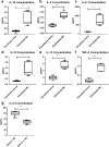
Macrophages impact inflammatory cytokine secretion in HSEs. Multiplex cytokine analysis for IL-1β (a), IL-2 (b), IL-8 (c), IL-10 (d), IL-13 (e), and TNF-A (f) showed significantly higher quantities in HSEs containing macrophages than those without. IL-6 levels (g) were significantly higher in HSEs containing DFUFs compared to those with NFFs, regardless of the presence of macrophages. N = 12. Mean ± SD. **p < 0.01; ***p < 0.001; ****p < 0.0001.
FIG. 6.
FIG. 6.
Diabetic monocytes secrete higher levels of inflammatory cytokines in HSEs. Blood monocytes from DFU (DM) or control subjects (CM) were incorporated into HSEs with DFUFs. (a–d) H&E staining indicated normal tissue development with monocytes from each subject incorporated. (e–h) Staining for HLADR indicated that monocytes differentiated into macrophages and expressed the HLADR marker. ELISA analysis for IL-1b (i), IL-6 (j), and IL-8 (k) showed significantly higher secretion levels in HSEs containing DM than CM, regardless of fibroblast source. Scale bars: 100 μm. Mean ± SD. n = 24. **** p < 0.0001. Color images are available online.

References

    1. Ramsey, S.D., Newton, K., Blough, D., et al. . Incidence, outcomes, and cost of foot ulcers in patients with diabetes. Diabetes Care 22, 382, 1999 - PubMed
    1. Boulton, A.J., Vileikyte, L., Ragnarson-Tennvall, G., and Apelqvist, J.. The global burden of diabetic foot disease. Lancet 366, 1719, 2005 - PubMed
    1. Pham, H.T., Economides, P.A., and Veves, A.. The role of endothelial function on the foot. Microcirculation and wound healing in patients with diabetes. Clin Podiatr Med Surg 15, 85, 1998 - PubMed
    1. Hesketh, M., Sahin, K.B., West, Z.E., and Murray, R.Z.. Macrophage phenotypes regulate scar formation and chronic wound healing. Int J Mol Sci 18, 2017 - PMC - PubMed
    1. Breslin, S., and O'Driscoll, L.. Three-dimensional cell culture: the missing link in drug discovery. Drug Discov Today 18, 240, 2013 - PubMed

Publication types

LinkOut - more resources