. 2020 Jan-Dec:16:1744806920975950.
doi: 10.1177/1744806920975950.

ASIC3 inhibition modulates inflammation-induced changes in the activity and sensitivity of Aδ and C fiber sensory neurons that innervate bone

Affiliations

ASIC3 inhibition modulates inflammation-induced changes in the activity and sensitivity of Aδ and C fiber sensory neurons that innervate bone

Michael Morgan et al. Mol Pain. 2020 Jan-Dec.

Abstract

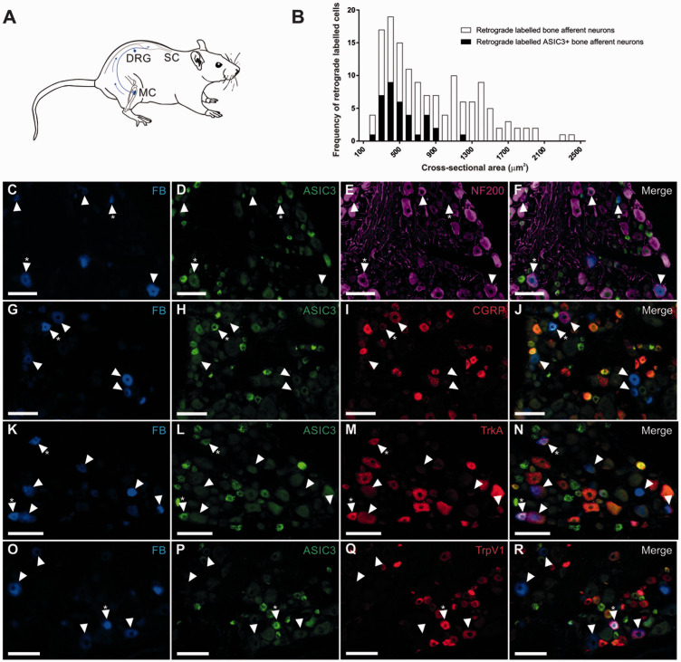
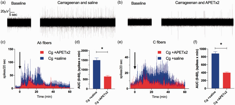
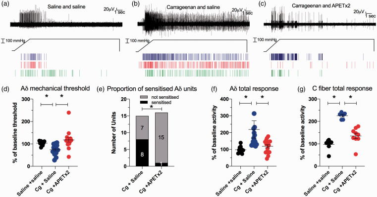
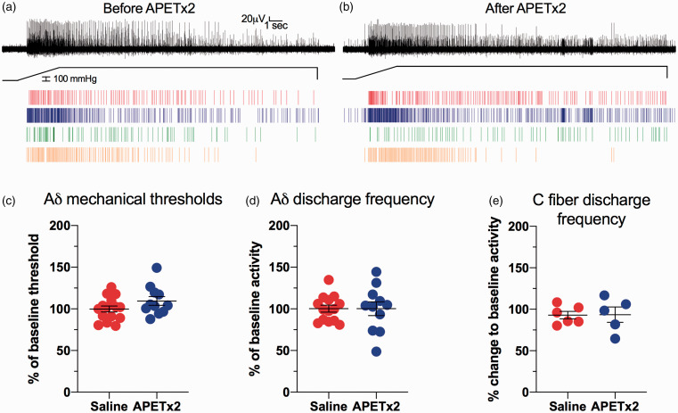
The Acid Sensing Ion Channel 3 (ASIC3) is a non-selective cation channel that is activated by acidification, and is known to have a role in regulating inflammatory pain. It has pro-algesic roles in a range of conditions that present with bone pain, but the mechanism for this has not yet been demonstrated. We aimed to determine if ASIC3 is expressed in Aδ and/or C fiber bone afferent neurons, and to explore its role in the activation and sensitization of bone afferent neurons after acute inflammation. A combination of retrograde tracing and immunohistochemistry was used to determine expression of ASIC3 in the soma of bone afferent neurons. A novel, in vivo, electrophysiological bone-nerve preparation was used to make recordings of the activity and sensitivity of bone afferent neurons in the presence of carrageenan-induced inflammation, with and without the selective ASIC3 inhibitor APET×2. A substantial proportion of bone afferent neurons express ASIC3, including unmyelinated (neurofilament poor) and small diameter myelinated (neurofilament rich) neurons that are likely to be C and Aδ nerve fibers respectively. Electrophysiological recordings revealed that application of APET×2 to the marrow cavity inhibited carrageenan-induced spontaneous activity of C and Aδ fiber bone afferent neurons. APET×2 also inhibited carrageenan-induced sensitization of Aδ and C fiber bone afferent neurons to mechanical stimulation, but had no effect on the sensitivity of bone afferent neurons in the absence of inflammation. This evidence supports a role for ASIC3 in the pathogenesis of pain associated with inflammation of the bone.

Keywords: ASIC3; Acid sensing ion channels; bone; electrophysiology; inflammatory pain; pain.


Conflict of interest statement

Declaration of Conflicting Interests: The author(s) declared no potential conflicts of interest with respect to the research, authorship, and/or publication of this article.

Figures

Figure 1.
Figure 1.
Images of retrograde labeled (Fast blue; FB) and immuno-labeled bone afferent neurons in sections through the DRG. (A) Schematic representation of the retrograde tracing approach used in this study. Fast Blue (FB) was injected into the marrow cavity (MC) of the rat tibia (N = 4). The tracer was taken up by nerve terminals and transported back to their soma in the dorsal root ganglion (DRG), permitting identification of sensory neurons that innervate the rat tibia. (B) Size/frequency distribution of all retrograde labeled bone afferent neurons analyzed in this study. Retrograde labeled bone afferent neurons that expressed ASIC3 were mostly small or medium sized neurons (<1800 μm2). (C)–(R) Images of retrograde and immuno-labeled bone afferent neurons in sections through the DRG. Each horizontal set of panels shows the same field of a single section. Arrowheads identify retrograde labeled bone afferent neurons throughout. Asterisks (*) indicate bone afferent neurons that are ASIC3+. C-F shows FB (C), ASIC3 immuno-labeling (D), NF200 immuno-labeling (E) and a merged image (F). G-J shows FB (G), ASIC3 immuno-labeling (H), CGRP immuno-labeling (I) and a merged image (J). K-N shows FB (K), ASIC3 immuno-labeling (L), TrkA immuno-labeling (M) and a merged image (N). O-R shows FB (O), ASIC3 immuno-labeling (P), TrpV1 immuno-labeling (Q) and a merged image (R). Scale bars = 100 µm.
Figure 2.
Figure 2.
Inhibition of ASIC3 with APETx2 inhibits carrageenan-induced activity in Aδ and C fiber bone afferent neurons. (a) and (b) are examples of whole-nerve recordings before (baseline) and after application of carrageenan+saline (a) or carrageen+APETx2 (b). These examples show a clear increase in spike activity when carrageenan is injected into the tibia in the presence of saline (a), but not when co-administered with APETx2 (b). (c) Frequency histograms of the total number of Aδ fiber spikes isolated from whole-nerve recordings before and after co-application of carrageenan with saline (N = 5) or APETx2 (1 µM; N = 6). (d) There was a reduction in the AUC0-60min for Aδ fiber spikes in animals treated with APETx2 compared to saline (Unpaired t-test, P < 0.001). (e) Frequency histograms of the total number of C fiber spikes isolated from whole-nerve recordings before and after co-application of carrageenan with saline (N = 4) or APETx2 (1 µM; N = 5). (f) There was a reduction in the AUC0-60min for C fiber spikes in animals treated with APETx2 compared to saline (Unpaired t-test, P < 0.001). Bin width = 20 seconds. Data are represented as mean ± SEM.
Figure 3.
Figure 3.
Inhibition of ASIC3 with APETx2 inhibits carrageenan-induced mechanical sensitization in Aδ and C fiber bone afferent neurons. (a)–(c) are examples of a whole-nerve recording and rasters of single Aδ fiber neuronal activity in response to a 300 mmHg ramp-and-hold pressure stimulus, in an animal injected with saline only (a), carrageenan+saline (b) and carrageenan+APETx2 (c). These examples show increased spike activity when carrageenan was injected into the tibia with saline (b), relative to when saline was injected into the tibia alone (a), and decreased spike activity when carrageenan was injected into the tibia in the presence of APETx2 (c), relative to when it was injected into the tibia in the presence of saline (b). (d) There was a reduction in threshold for activation of single Aδ fiber units in animals administered carrageenan+saline (N = 7, n = 15), relative to those administered saline alone (N = 7, n = 10) (Dunnett test, P < 0.05), and an increase in the threshold for activation in animals administered carrageenan+APETx2 (N = 7, n = 16), relative to those administered carrageenan+saline (Dunnett test, P < 0.05). (e) Significantly fewer of the single units were sensitized in animals treated with carrageenan+APETx2 compared to carrageenan+saline (Chi-square test, P < 0.05). (f) There was an increase in single Aδ fiber unit discharge frequency in animals administered carrageenan+saline (N = 7, n = 15), relative to those administered saline alone (N = 7, n = 10) (Dunnett test, P < 0.05), and a reduction in discharge frequency in animals administered carrageenan+APETx2 (N = 7, n = 16), relative to those administered carrageenan+saline (Dunnett test, P < 0.05). (g) There was an increase in C fiber discharge in animals treated with carrageenan+saline (N = 6) relative to animals treated with saline alone (N = 7) (Dunn’s test; P < 0.05), and reduced C fiber activity for animals treated with carrageenan+APETx2 (N = 9) relative to animals treated with carrageenan+saline (Dunn’s test, P < 0.05).
Figure 4.
Figure 4.
Inhibition of ASIC3 with APETx2 does not affect mechanical sensitivity of Aδ and C fiber bone afferent neurons. (a) and (b) are examples of a whole-nerve recording and rasters of single Aδ fiber activity in response to a 300 mmHg ramp-and-hold pressure stimulus, in an animal before (a) and after (b) injection of APETx2. They show no difference in spike activity before (a) or after (b) injection of APETx2. (c) There was no change to threshold for activation of single Aδ fiber units in animals administered APETx2 (N = 5, n = 11), relative to those administered saline alone (N = 7, n = 17) (nested t-test, P > 0.05). (d) There was no change to discharge frequency in animals administered APETx2 (N = 5, n = 11), relative to those administered saline alone (N = 7, n = 17) (nested t-test, P > 0.05). (f) There was no change to whole-nerve C fiber discharge in animals treated with APETx2 (N = 5), relative to animals treated with saline alone (N = 6) (Mann Whitney test; P > 0.05).

Similar articles

Cited by

References

    1. Berenbaum F. Osteoarthritis as an inflammatory disease (osteoarthritis is not osteoarthrosis!). Osteoarth Cartil 2013; 21: 16–21. - PubMed
    1. Bove SE, Flatters SJ, Inglis JJ, Mantyh PW. New advances in musculoskeletal pain. Brain Res Rev 2009; 60: 187–201. - PMC - PubMed
    1. Haegerstam GA. Pathophysiology of bone pain: a review. Acta Orthop Scand 2001; 72: 308–317. - PubMed
    1. Mamet J, Baron A, Lazdunski M, Voilley N. Proinflammatory mediators, stimulators of sensory neuron excitability via the expression of acid-sensing ion channels. J Neurosci 2002; 22: 10662–10670. - PMC - PubMed
    1. Nagae M, Hiraga T, Wakabayashi H, Wang L, Iwata K, Yoneda T. Osteoclasts play a part in pain due to the inflammation adjacent to bone. Bone 2006; 39: 1107–1115. - PubMed

Publication types

MeSH terms

LinkOut - more resources