. 2020 Dec 7;22(1):83.
doi: 10.1186/s12968-020-00684-2.

Clinical validation of three cardiovascular magnetic resonance techniques to measure strain and torsion in patients with suspected coronary artery disease

Affiliations

Clinical validation of three cardiovascular magnetic resonance techniques to measure strain and torsion in patients with suspected coronary artery disease

Johan Kihlberg et al. J Cardiovasc Magn Reson. .

Abstract

Background: Several cardiovascular magnetic resonance (CMR) techniques can measure myocardial strain and torsion with high accuracy. The purpose of this study was to compare displacement encoding with stimulated echoes (DENSE), tagging and feature tracking (FT) for measuring circumferential and radial myocardial strain and myocardial torsion in order to assess myocardial function and infarct scar burden both at a global and at a segmental level.

Method: 116 patients with a high likelihood of coronary artery disease (European SCORE > 15%) underwent CMR examination including cine images, tagging, DENSE and late gadolinium enhancement (LGE) in the short axis direction. In total, 97 patients had signs of myocardial disease and 19 had no abnormalities in terms of left ventricular (LV) wall mass index, LV ejection fraction, wall motion, LGE or a history of myocardial infarction. Thirty-four patients had myocardial infarct scar with a transmural LGE extent (transmurality) that exceeded 50% of the wall thickness in at least one segment. Global circumferential strain (GCS) and global radial strain (GRS) was analyzed using FT of cine loops, deformation of tag lines or DENSE displacement.

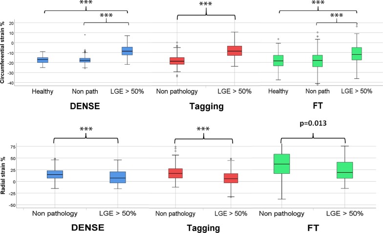
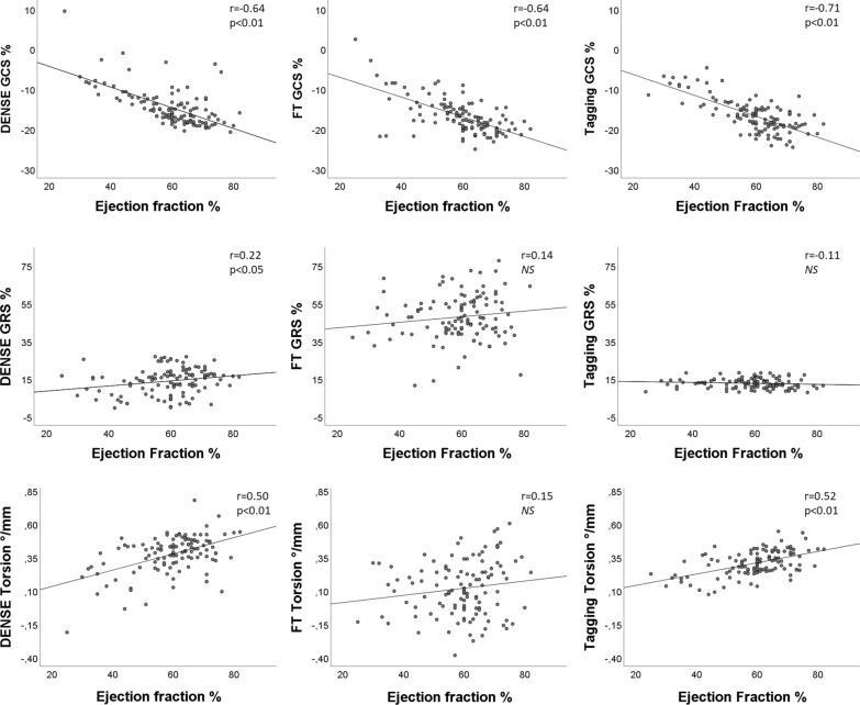
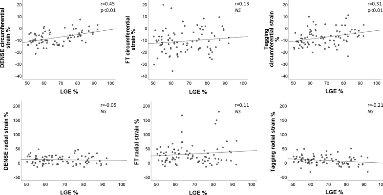
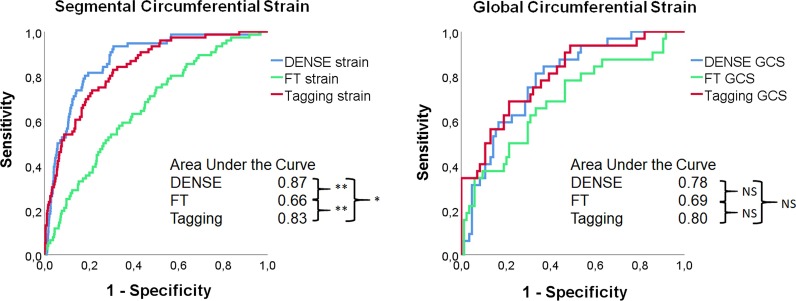
Results: DENSE and tagging both showed high sensitivity (82% and 71%) at a specificity of 80% for the detection of segments with > 50% LGE transmurality, and receiver operating characteristics (ROC) analysis showed significantly higher area under the curve-values (AUC) for DENSE (0.87) than for tagging (0.83, p < 0.001) and FT (0.66, p = 0.003). GCS correlated with global LGE when determined with DENSE (r = 0.41), tagging (r = 0.37) and FT (r = 0.15). GRS had a low but significant negative correlation with LGE; DENSE r = - 0.10, FT r = - 0.07 and tagging r = - 0.16. Torsion from DENSE and tagging had a weak correlation (- 0.20 and - 0.22 respectively) with global LGE.

Conclusion: Circumferential strain from DENSE detected segments with > 50% scar with a higher AUC than strain determined from tagging and FT at a segmental level. GCS and torsion computed from DENSE and tagging showed similar correlation with global scar size, while when computed from FT, the correlation was lower.

Keywords: DENSE; Feature tracking; Myocardial infarction; Strain; Tagging; Torsion.


Conflict of interest statement

The authors declare that they have no competing interests.

Figures

Fig. 1
Fig. 1
Strain in segments with scar transmurality > 50%. Boxplot of segmental circumferential strain (top row) and radial strain (lower row) assessed using three techniques in ten healthy subjects (160 segments) and patients with no signs of pathology in terms of left ventricular (LV) wall mass index, systolic blood pressure (SBP), diastolic blood pressure (DBP), wall motion, late gadolinium enhancement (LGE) and a history of myocardial infarction, (“Non pathology”, 304 segments) and segments with LGE in excess of 50% wall thickness (LGE > 50%, 84 segments). The box plots show median, the two central quartiles in the box, one quartile in each whisker and outliers. Student’s t-test showed significant differences between the two patient groups for all three techniques.***p < 0.001. Healthy subjects depicted for comparison
Fig. 2
Fig. 2
Anteroseptal infarct scar. Images and measurements in a 68-year-old male with extensive anteroseptal and apical infarct scar with transmurality > 50%. Scar volume was 25% of the LV myocardial mass, and the LV ejection fraction (EF) was 42%. The left-hand panel LGE images (ad) show the extent of scar. The right-hand panel bull’s eye (h) shows percentages for segmental transmurality. The remaining panels show circumferential strain assessed by (e) FT, (f) Tagging, (g) DENSE
Fig. 3
Fig. 3
Correlation of global strain and torsion with left ventricular ejection fraction. Pearson correlations (r) between global circumferential strain (GCS, top row), global radial strain (GRS, middle row) and torsion (lower row) and left ventricular ejection fraction. GCR, GRS and torsion assessed by DENSE, FT, and tagging for 116 patients plotted against ejection fraction. The best-fit linear regression as well as the Pearson correlation coefficient and its p-value are shown for each plot. NS no significant correlation
Fig. 4
Fig. 4
Correlation of global strain and torsion with late gadolinium enhancement (LGE). Pearson correlations (r) between global circumferential strain (GCS, top row), global radial strain (GRS, middle row) and torsion (lower row) vs global LGE as a percentage of the total wall mass. GCR, GRS and torsion assessed by DENSE, FT, and tagging for 116 patients plotted against LGE. The best-fit linear regression as well as the Pearson correlation coefficient and its p-value are shown for each plot. NS no significant correlation. The best correlation is seen between tagging strain amplitude and LGE while no correlation is seen between torsion and LGE
Fig. 5
Fig. 5
Segmental strain in segments with late gadolinium enhancement (LGE) transmurality > 50%. Relationship between segmental circumferential strain (top row) and segmental radial strain (lower row) and LGE transmurality in segments with LGE > 50% (84 segments from 34 patients). Strain was assessed by DENSE, FT and tagging. The best-fit linear regression as well as the Pearson correlation coefficient and its p-value are shown for each plot. NS no significant correlation
Fig. 6
Fig. 6
Receiver operator characteristics (ROC)-analysis of segmental and global strain. To the left ROC curves for the detection of infarcted segments with scar transmurality exceeding 50% LGE (84 [34 patients of 116] out of 1856 segments) using circumferential strain from DENSE, FT and Tagging at end systole. To the right ROC curves for the detection of infarcted segments with scar transmurality exceeding 50% LGE (34 patients out of 116 patients) using global circumferential strain (GCS) from DENSE, FT and Tagging at end systole. AUC values are shown in the lower right corner with significance level *p < 0.05 and **p < 0.01

References

    1. Fratz S, Chung T, Greil GF, Samyn MM, Taylor AM, Valsangiacomo Buechel ER, et al. Guidelines and protocols for cardiovascular magnetic resonance in children and adults with congenital heart disease: SCMR expert consensus group on congenital heart disease. J Cardiovasc Magn Reson. 2013;15(1):1–26. doi: 10.1186/1532-429X-15-51. - DOI - PMC - PubMed
    1. Mirsky I, Parmley WW. Assessment of passive elastic stiffness for isolated heart muscle and the intact heart. Circ Res. 1973;33(2):233–243. doi: 10.1161/01.RES.33.2.233. - DOI - PubMed
    1. Henson RE, Song SK, Pastorek JS, Ackerman JJH, Lorenz CH. Left ventricular torsion is equal in mice and humans. Am J Physiol. 2000;278(4):H1117–H1123. - PubMed
    1. Codreanu I, Robson MD, Golding SJ, Jung BA, Clarke K, Holloway CJ. Longitudinally and circumferentially directed movements of the left ventricle studied by cardiovascular magnetic resonance phase contrast velocity mapping. J Cardiovasc Magn Reson. 2010;12(1):1–8. doi: 10.1186/1532-429X-12-48. - DOI - PMC - PubMed
    1. Osman NF, Kerwin WS, McVeigh ER, Prince JL. Cardiac motion tracking using CINE harmonic phase (HARP) magnetic resonance imaging. Magn Reson Med. 1999;42(6):1048–1060. doi: 10.1002/(SICI)1522-2594(199912)42:6<1048::AID-MRM9>3.0.CO;2-M. - DOI - PMC - PubMed

Publication types

MeSH terms