

Advanced 3D "Modeling" and "Printing" for the Surgical Planning of a Successful Case of Thoraco-Omphalopagus Conjoined Twins Separation
- PMID: 33281611
- PMCID: PMC7691583
- DOI: 10.3389/fphys.2020.566766
Advanced 3D "Modeling" and "Printing" for the Surgical Planning of a Successful Case of Thoraco-Omphalopagus Conjoined Twins Separation
Abstract
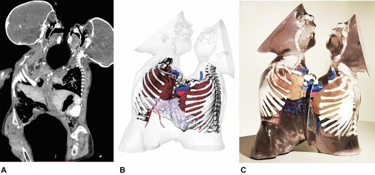
Background: The surgical separation of two Conjoined Twins is a particularly complex operation. Surgical times are particularly long and post-operative complications are very frequent in this type of procedure. We report a clinical case of surgical separation of two thoraco-omphalopagus conjoined twins in which, thanks to the use of (3D) three dimensional technologies, we were able to significantly reduce operative times and improve clinical outcomes.
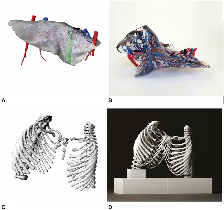
Methods: We performed a 3D reconstruction of the anatomical parts involved in congenital fusion using Computer Tomography (CT) images.We obtained virtual anatomical models of the patients which allowed us to estimate essential details as the residual post-operative thoracic volume as well as the exact position of resection planes for both the general separation and for the hepatic splitting procedure. Subsequently, we printed 3D anatomical models of the thoracic cage and sternum and of the liver with the plane of resection. Finally, we printed an additional 3D anatomical model of the two patients representing different organs with multiple colors and materials.
Results: The use of 3D printing reduced the duration of surgery by 30% with a favorable patient outcome. Two years after the operation, the patients do not present any type of deficit and have a normal life without any significant complication.
Conclusion: Virtual anatomical 3D models and 3D printing represent a valid technological tool to support complex surgical operations, especially in pre-surgical planning. 3D models are important tools to better understand complex anatomy and to discuss clinical cases among members of the surgical team.
Keywords: 3D images; 3D printing; 3D printing in cardiothoracic surgery; 3D printing in surgery; conjoined twins.
Copyright © 2020 Inserra, Borro, Spada, Frediani and Secinaro.
Figures
Similar articles
-
Hepatic separation of conjoined twins: Operative technique and review of three-dimensional model utilization.J Pediatr Surg. 2020 Dec;55(12):2828-2835. doi: 10.1016/j.jpedsurg.2020.06.047. Epub 2020 Jul 15. J Pediatr Surg. 2020. PMID: 32792165 Review.
-
Successful Surgical Separation of Thoraco-Omphalopagus Symmetrical Conjoined Twins in Iran: Two Case Reports.Iran J Med Sci. 2020 Mar;45(2):143-147. doi: 10.30476/ijms.2019.81060.. Iran J Med Sci. 2020. PMID: 32210492 Free PMC article.
-
Conjoined Twin Separation: Integration of Three-Dimensional Modeling for Optimization of Surgical Planning.J Craniofac Surg. 2017 Jan;28(1):4-10. doi: 10.1097/SCS.0000000000003412. J Craniofac Surg. 2017. PMID: 27977489
-
Use of local perforator flaps for closure of a thoraco-omphalopagus conjoined twin defect after separation during the COVID-19 pandemic.Heliyon. 2021 Jul;7(7):e07443. doi: 10.1016/j.heliyon.2021.e07443. Epub 2021 Jun 30. Heliyon. 2021. PMID: 34226881 Free PMC article.
-
The clinical use of 3D printing in surgery.Updates Surg. 2018 Sep;70(3):381-388. doi: 10.1007/s13304-018-0586-5. Epub 2018 Aug 30. Updates Surg. 2018. PMID: 30167991 Review.
Cited by
-
Postnatal imaging of conjoined twins: a customized multimodality approach.Pediatr Radiol. 2023 Oct;53(11):2291-2304. doi: 10.1007/s00247-023-05709-3. Epub 2023 Jul 19. Pediatr Radiol. 2023. PMID: 37466734 Free PMC article. Review.
-
Advancements and applications of 3D printing in pediatric orthopedics: A comprehensive review.J Child Orthop. 2025 Mar 15;19(2):119-138. doi: 10.1177/18632521251318552. eCollection 2025 Apr. J Child Orthop. 2025. PMID: 40098806 Free PMC article. Review.
-
3D Printing in a hospital: Centralized clinical implementation and applications for comprehensive care.Digit Health. 2023 Dec 20;9:20552076231221899. doi: 10.1177/20552076231221899. eCollection 2023 Jan-Dec. Digit Health. 2023. PMID: 38130801 Free PMC article.
-
The Role of Three-dimensional Printed Models in Women's Health.Womens Health (Lond). 2023 Jan-Dec;19:17455057231199040. doi: 10.1177/17455057231199040. Womens Health (Lond). 2023. PMID: 37688305 Free PMC article. Review.
References
-
- Bugaje M. A., McHoney M., Ameh E. A., Lakhoo K. (2010). “Omphalitis,” in Paediatric Surgery: A Comprehensive Text For Africa. help_pedsurgeryafricavolume01.pdf, Vol. 1 eds Stephen E. A., Bickler W., Lakhoo K., Poenaru D. (Seattle, Washington, DC: Global Help; ), 124–128.
-
- Jensen K., Bjerrum F., Hansen H. J., Petersen R. H., Pedersen J. H., Konge L. (2015). A new possibility in thoracoscopic virtual reality simulation training: development and testing of a novel virtual reality simulator for video-assisted thoracoscopic surgery lobectomy. Interact. Cardiovasc. Thorac. Surg. 21 420–426. 10.1093/icvts/ivv183 - DOI - PubMed
LinkOut - more resources
Full Text Sources