. 2020 Dec;54(6):319-323.
doi: 10.1007/s13139-020-00673-4. Epub 2020 Nov 9.

Isolated Testicular Metastasis from Prostate Cancer Detected on Ga-68 PSMA PET/CT Scan

Affiliations

Isolated Testicular Metastasis from Prostate Cancer Detected on Ga-68 PSMA PET/CT Scan

Nitin Gupta et al. Nucl Med Mol Imaging. 2020 Dec.

Abstract

Although prostate cancer can metastasize to any part of the body, isolated testicular metastasis is very rare and only few cases have been reported so far. Here we present a case of 65-year-old male patient, known case of prostate adenocarcinoma, referred for 68Ga-PSMA PET/CT scan, post radiotherapy, and androgen deprivation therapy, for rising serum PSA levels. He was found to have an isolated testicular metastasis on the scan. This report highlights the importance of 68Ga-PSMA PET-CT scan in detecting these unusual and rare sites of metastasis from prostate cancer.

Keywords: 68Ga-PSMA PET/CT; Adenocarcinoma prostate; Testicular metastasis.


Conflict of interest statement

Conflict of InterestDr. Nitin Gupta, Dr. Sudip Dey, Dr. Ritu Verma, and Dr. Ethel S.Belho declare that they have no conflicts of interest. There is no source of funding.

Figures
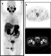
Fig. 1
Fig. 1
A 65-year-old male patient, known case of adenocarcinoma prostate gland (Gleason’s score 7 (4 + 3)), diagnosed 3 years ago, post radiotherapy and androgen deprivation therapy, presented with rising serum PSA levels, with a recent value of 7.07 ng/ml was referred for 68Ga-PSMA PET/CT scan. Maximal intensity projection (MIP) of 68Ga-PSMA PET/CT scan (a) and axial PET, CT, and fused PET/CT images (bd) showed mild inhomogeneous PSMA uptake in the prostate gland. Axial and Sagittal PET, CT, and fused PET/CT images (ej) revealed PSMA avid ill-defined heterogeneously enhancing lesions in the right testis likely metastatic. Later, the patient underwent bilateral orchiectomy, and histopathologic report confirmed metastatic prostatic adenocarcinoma to the right testis. Prostate-specific membrane antigen is a transmembrane protein, which is overexpressed in prostate carcinoma cells [1]. 68Ga-PSMA ligand PET/CT is a highly sensitive and specific imaging technique for the detection of primary and metastatic prostate cancer [2]. Metastatic prostate cancer lesions are commonly found in lymph nodes, bones, and lungs but infrequently seen in testes [3]. Several pathophysiological mechanisms have been described to explain metastatic spread to the testes, like retrograde venous extension, retrograde lymphatic spread, or direct extension through the vas deferens. Usually, testicular metastases in prostate cancer are suggestive of advanced prostate disease. Very rarely, the testis may be the isolated site of metastatic spread, as seen in our case. To the best of our knowledge, very few case reports have been published about solitary testicular metastasis detected by 68Ga-PSMA PET/CT scan [–7]. However, in literature, there are few articles that describe 68Ga-PSMA uptake in non-prostatic conditions [–10]. Metastatic disease to testicles from solid tumors is very rare; a study of 738 patients with solid malignant neoplasms found metastasis to testicles in 0.68% of patients [11]. Approximately 4% of testicular metastases are found incidentally during orchiectomy for advanced prostate cancer [12]. This report highlights the rare finding of testicular metastasis from prostate cancer and the potential of 68Ga-PSMA-targeted PET/CT scan for detection of metastases even in these atypical sites
Fig. 1
Fig. 1
A 65-year-old male patient, known case of adenocarcinoma prostate gland (Gleason’s score 7 (4 + 3)), diagnosed 3 years ago, post radiotherapy and androgen deprivation therapy, presented with rising serum PSA levels, with a recent value of 7.07 ng/ml was referred for 68Ga-PSMA PET/CT scan. Maximal intensity projection (MIP) of 68Ga-PSMA PET/CT scan (a) and axial PET, CT, and fused PET/CT images (bd) showed mild inhomogeneous PSMA uptake in the prostate gland. Axial and Sagittal PET, CT, and fused PET/CT images (ej) revealed PSMA avid ill-defined heterogeneously enhancing lesions in the right testis likely metastatic. Later, the patient underwent bilateral orchiectomy, and histopathologic report confirmed metastatic prostatic adenocarcinoma to the right testis. Prostate-specific membrane antigen is a transmembrane protein, which is overexpressed in prostate carcinoma cells [1]. 68Ga-PSMA ligand PET/CT is a highly sensitive and specific imaging technique for the detection of primary and metastatic prostate cancer [2]. Metastatic prostate cancer lesions are commonly found in lymph nodes, bones, and lungs but infrequently seen in testes [3]. Several pathophysiological mechanisms have been described to explain metastatic spread to the testes, like retrograde venous extension, retrograde lymphatic spread, or direct extension through the vas deferens. Usually, testicular metastases in prostate cancer are suggestive of advanced prostate disease. Very rarely, the testis may be the isolated site of metastatic spread, as seen in our case. To the best of our knowledge, very few case reports have been published about solitary testicular metastasis detected by 68Ga-PSMA PET/CT scan [–7]. However, in literature, there are few articles that describe 68Ga-PSMA uptake in non-prostatic conditions [–10]. Metastatic disease to testicles from solid tumors is very rare; a study of 738 patients with solid malignant neoplasms found metastasis to testicles in 0.68% of patients [11]. Approximately 4% of testicular metastases are found incidentally during orchiectomy for advanced prostate cancer [12]. This report highlights the rare finding of testicular metastasis from prostate cancer and the potential of 68Ga-PSMA-targeted PET/CT scan for detection of metastases even in these atypical sites

Similar articles

Cited by

References

    1. Silver DA, Pellicer I, Fair WR, et al. Prostate-specific membrane antigen expression in normal and malignant human tissues. Clin Cancer Res. 1997;3:81–85. - PubMed
    1. Eiber M, Maurer T, Souvatzoglou M, et al. Evaluation of hybrid 68Ga-PSMA ligand PET/CT in 248 patients with biochemical recurrence after radical prostatectomy. J Nucl Med. 2015;56:668–674. doi: 10.2967/jnumed.115.154153. - DOI - PubMed
    1. Bubendorf L, Schöpfer A, Wagner U, et al. Metastatic patterns of prostate cancer: an autopsy study of 1,589 patients. Hum Pathol. 2000;31:578–583. doi: 10.1053/hp.2000.6698. - DOI - PubMed
    1. da Cunha ML, de Oliveira Rodrigues C, de Araújo MPL, de Freitas Junior CH, Ferrigno R. Solitary testicular metastasis from prostate cancer. A case report diagnosed by PET/CT with PSMA. Eur J Nucl Med Mol Imaging. 2018;45:888–889. doi: 10.1007/s00259-018-3942-2. - DOI - PubMed
    1. Deeb IA, Khdhir MA, Bulbul M, Haidar M. Solitary metastasis of prostatic adenocarcinoma to the testicle detected by 68Gaprostate-specific membrane antigen positron emission tomography/computed tomography. Indian J Nucl Med. 2020;35:61–62. doi: 10.4103/ijnm.IJNM_182_19. - DOI - PMC - PubMed

LinkOut - more resources