Review
. 2020 Nov;12(11):6954-6965.
doi: 10.21037/jtd-2019-cptn-03.

Putting artificial intelligence (AI) on the spot: machine learning evaluation of pulmonary nodules

Affiliations
Review

Putting artificial intelligence (AI) on the spot: machine learning evaluation of pulmonary nodules

Yasmeen K Tandon et al. J Thorac Dis. 2020 Nov.

Abstract

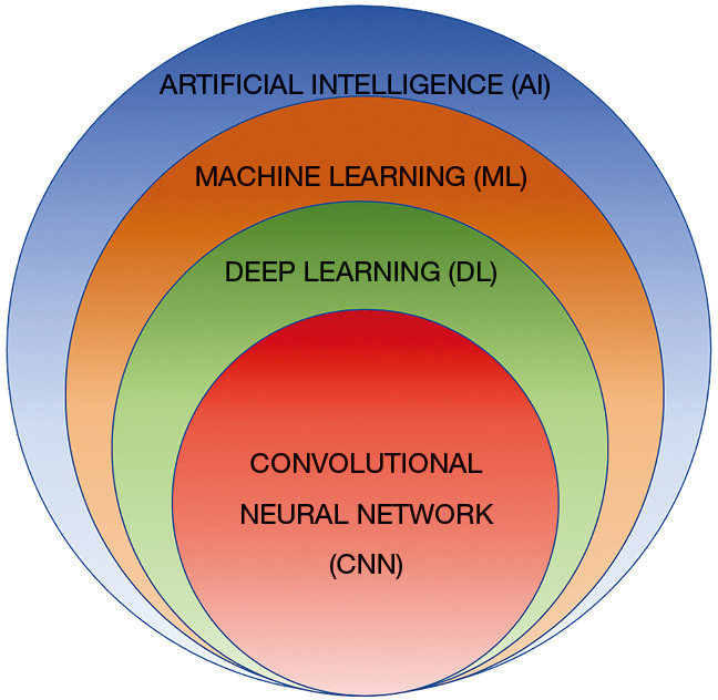
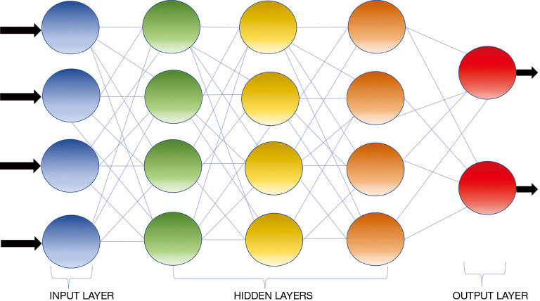
Lung cancer remains the leading cause of cancer related death world-wide despite advances in treatment. This largely relates to the fact that many of these patients already have advanced diseases at the time of initial diagnosis. As most lung cancers present as nodules initially, an accurate classification of pulmonary nodules as early lung cancers is critical to reducing lung cancer morbidity and mortality. There have been significant recent advances in artificial intelligence (AI) for lung nodule evaluation. Deep learning (DL) and convolutional neural networks (CNNs) have shown promising results in pulmonary nodule detection and have also excelled in segmentation and classification of pulmonary nodules. This review aims to provide an overview of progress that has been made in AI recently for pulmonary nodule detection and characterization with the ultimate goal of lung cancer prediction and classification while outlining some of the pitfalls and challenges that remain to bring such advancements to routine clinical use.

Keywords: Artificial intelligence (AI); machine learning (ML); pulmonary nodule.


Conflict of interest statement

Conflicts of Interest: The authors have completed the ICMJE uniform disclosure form (available at: http://dx.doi.org/10.21037/jtd-2019-cptn-03). The series “Contemporary Practice in Thoracic Neoplasm Diagnosis, Evaluation and Treatment” was commissioned by the editorial office without any funding or sponsorship. CWK served as the unpaid Guest Editor of the series and serves as an unpaid editorial board member of Journal of Thoracic Disease from Dec 2018 to Nov 2020. Dr. BJB reports personal fees from Promedior, LLC, other royalties from Imbio, LLC, outside the submitted work. In addition, Dr. BJB has a patent SYSTEMS AND METHODS FOR ANALYZING IN VIVO TISSUE VOLUMES USING MEDICAL IMAGING pending and intellectual property rights to CANARY software but no financial relationships from that software. The authors have no other conflicts of interest to declare.

Figures

Figure 1
Figure 1
Euler diagram demonstrating the AI hierarchy. ML is a subfield of AI where algorithms can recognize and learn patterns using complex data sets to produce without explicit programming. DL can be conceptualized as a class of ML where algorithms are organized into many processing layers based on artificial neural networks, similar to the human brain. CNN is the type of DL model most commonly used for medical imaging presently. AI, artificial intelligence; ML, machine learning; DL, deep learning; CNN, convolutional neural network.
Figure 2
Figure 2
Schematic representation of a convolutional neural network containing an input layer, three hidden connected layers, and an output layer. The computational strength of such network lies in the integration of multiple “neurons” (represented by the circles) within the deep hidden layers between the input and output layers. Typically the outputs of one layer serve as the input of the next layer.
Figure 3
Figure 3
General illustration of a feature based machine learning nodule classification and risk stratification model [modified from Computer-Aided Nodule Assessment and Risk Yield (CANARY), Mayo Clinic, Rochester, MN, USA]. (A) Nodule Classification. After nodule segmentation, radiomic features are extracted from the images. Following feature-pathology correlation and feature selection, the machine learning model is trained to classify pulmonary nodules. There is typically a validation step with a separate set of data to further refine the algorithm before final testing prior to use. (B) Risk stratification. Multiple nodules with features representative of the population and the corresponding survival data are used to train the algorithm, which in turn classifies the features into three main groups in the case of CANARY and performs survival analysis of each group of nodules (the orange group has good, the green group has intermediate and the purple group has poor prognosis).

References

    1. Siegel RL, Miller KD, Jemal A. Cancer statistics, 2019. CA Cancer J Clin 2019;69:7-34. 10.3322/caac.21551 - DOI - PubMed
    1. Kligerman S, Cai L, White CS. The effect of computer-aided detection on radiologist performance in the detection of lung cancers previously missed on a chest radiograph. J Thorac Imaging 2013;28:244-52. 10.1097/RTI.0b013e31826c29ec - DOI - PubMed
    1. de Hoop B, De Boo DW, Gietema HA, et al. Computer-aided detection of lung cancer on chest radiographs: effect on observer performance. Radiology 2010;257:532-40. 10.1148/radiol.10092437 - DOI - PubMed
    1. Schalekamp S, van Ginneken B, Koedam E, et al. Computer-aided detection improves detection of pulmonary nodules in chest radiographs beyond the support by bone-suppressed images. Radiology 2014;272:252-61. 10.1148/radiol.14131315 - DOI - PubMed
    1. Li F, Arimura H, Suzuki K, et al. Computer-aided detection of peripheral lung cancers missed at CT: ROC analyses without and with localization. Radiology 2005;237:684-90. 10.1148/radiol.2372041555 - DOI - PubMed