Review
. 2020 Nov;12(11):6978-6991.
doi: 10.21037/jtd-2019-cptn-09.

Thoracic positron emission tomography: 18F-fluorodeoxyglucose and beyond

Affiliations
Review

Thoracic positron emission tomography: 18F-fluorodeoxyglucose and beyond

Timothy J Jaykel et al. J Thorac Dis. 2020 Nov.

Abstract

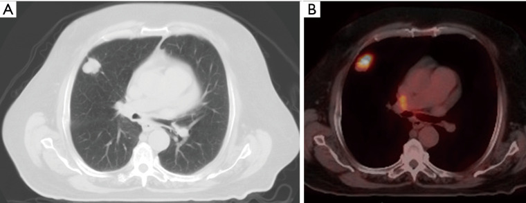
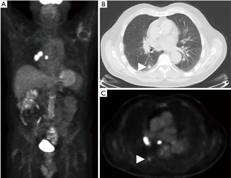
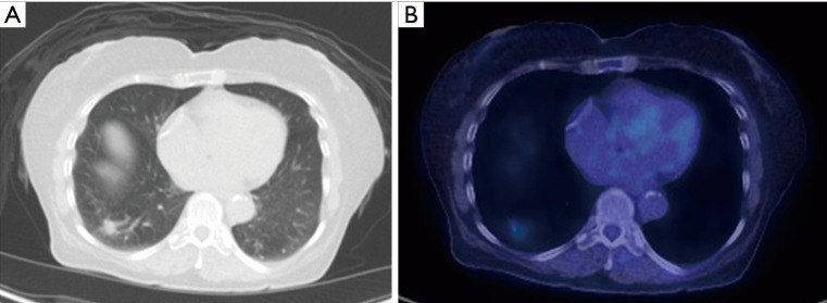
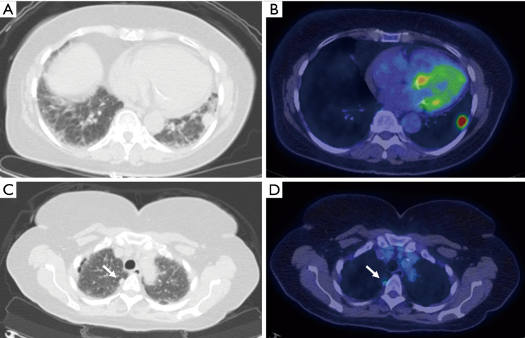
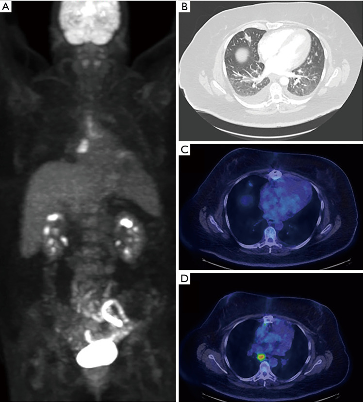
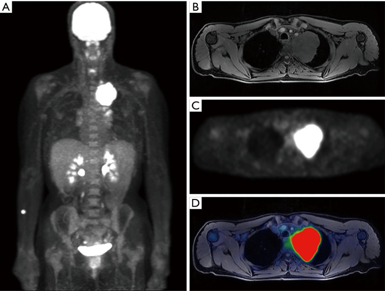
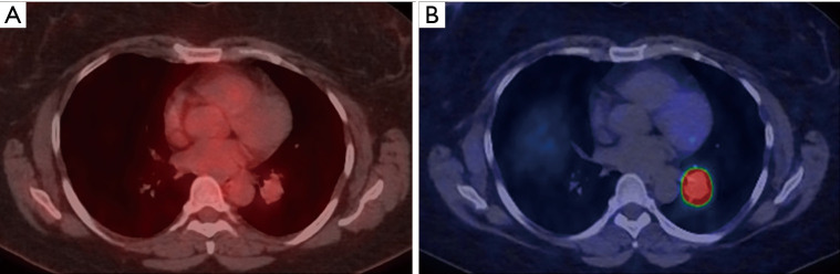
Ongoing technologic and therapeutic advancements in medicine are now testing the limits of conventional anatomic imaging techniques. The ability to image physiology, rather than simply anatomy, is critical in the management of multiple disease processes, especially in oncology. Nuclear medicine has assumed a leading role in detecting, diagnosing, staging and assessing treatment response of various pathologic entities, and appears well positioned to do so into the future. When combined with computed tomography (CT) or magnetic resonance imaging (MRI), positron emission tomography (PET) has become the sine quo non technique of evaluating most solid tumors especially in the thorax. PET/CT serves as a key imaging modality in the initial evaluation of pulmonary nodules, often obviating the need for more invasive testing. PET/CT is essential to staging and restaging in bronchogenic carcinoma and offers key physiologic information with regard to treatment response. A more recent development, PET/MRI, shows promise in several specific lung cancer applications as well. Additional recent advancements in the field have allowed PET to expand beyond imaging with 18F-flurodeoxyglucose (FDG) alone, now with the ability to specifically image certain types of cell surface receptors. In the thorax this predominantly includes 68Ga-DOTATATE which targets the somatostatin receptors abundantly expressed in neuroendocrine tumors, including bronchial carcinoid. This receptor targeted imaging technique permits targeting these tumors with therapeutic analogues such as 177Lu labeled DOTATATE. Overall, the proper utilization of PET in the thorax has the ability to directly impact and improve patient care.

Keywords: Positron emission tomography (PET); bronchogenic carcinoma; positron emission tomography/computed tomography (PET/CT); positron emission tomography/magnetic resonance imaging (PET/MRI); pulmonary nodule.


Conflict of interest statement

Conflicts of Interest: All authors have completed the ICMJE uniform disclosure form (available at: http://dx.doi.org/10.21037/jtd-2019-cptn-09). The series “Contemporary Practice in Thoracic Neoplasm Diagnosis, Evaluation and Treatment” was commissioned by the editorial office without any funding or sponsorship. The authors have no other conflicts of interest to declare.

Figures

Figure 1
Figure 1
A 74-year-old man found to have a pulmonary nodule at chest radiograph (not shown). Axial CT image (A) shows a nodule in the right upper lobe with indistinct margins. Fused axial images from 18F-FDG PET/CT (B) show marked FDG uptake within this lesion, highly suspicious for carcinoma. The lesion ultimately underwent biopsy, confirming lung adenocarcinoma.
Figure 2
Figure 2
A 78-year-old female with a right lower lobe pulmonary nodule, incidentally seen on CT to assess aortic dissection repair, was further characterized with FDG PET/CT (A). Axial CT (B) and PET (C) images demonstrate a mildly FDG avid right lower lobe pulmonary nodule (arrowheads, SUVmax 2.2) and markedly FDG avid right hilar lymphadenopathy (SUVmax 13.6). Discordant degree of uptake within the nodule versus the draining lymphadenopathy is most consistent with an infectious etiology, specifically chronic fungal infection (so called “flip-flop-fungus” sign). Serological evaluation was positive for histoplasmosis.
Figure 3
Figure 3
A 69-year-old man with a previous metastatic malignancy with enlarging right lower lobe pulmonary nodule (A). Fused axial images from 18F-FDG PET/CT (B) demonstrate minimal FDG uptake. The pulmonary nodule was biopsied and was negative for malignancy but was consistent for focal organizing pneumonia.
Figure 4
Figure 4
A 68-year-old man with metastatic squamous cell carcinoma presents for restaging. Axial fused 18F-FDG PET/CT demonstrates an FDG avid right middle lobe primary tumor and a large left pleural effusion without uptake. Laboratory testing of the thoracentesis was negative for malignancy.
Figure 5
Figure 5
A 74-year-old male presenting with shortness of breath and fatigue. MIP reconstruction from FDG PET (A) shows a large FDG avid left small cell lung cancer with numerous additional foci of abnormal uptake throughout the body. Fused axial PET/CT images reveal that bilateral hilar lymph nodes (B), vertebral body (C), liver (D), and the adrenals (E) have metastatic disease.
Figure 6
Figure 6
A 74-year-old man status post right pneumonectomy for squamous cell carcinoma was imaged for surveillance. Axial CT (A) and fused FDG PET/CT (B) images show mild uptake in the left hilum (arrows). Follow-up scan obtained 4 months later demonstrated unchanged size (C) but interval increase FDG uptake in the hilar mass (D), suspicious for recurrent disease.
Figure 7
Figure 7
A 74-year-old woman with biopsy proven left lower lobe bronchogenic carcinoma. Index lesion is shown on axial CT image (A) with increased FDG uptake (B). Smaller right upper lobe pulmonary nodule (arrows) on axial CT (C) also demonstrates mild increased FDG uptake (D). This nodule was biopsied and confirmed to represent metastatic deposit.
Figure 8
Figure 8
A 73-year-old female presenting for initial staging of NSCL demonstrating the utility of FDG PET/CT. MIP reconstruction (A) demonstrates widespread metastatic disease. Axial fused PET/CT images show: hypermetabolic right brain metastasis (B), hypermetabolic primary right upper lobe malignancy (C), right pleural metastasis (D) and right iliac bone metastasis (E).
Figure 9
Figure 9
A 58-year-old female with incidentally discovered right middle lobe pulmonary nodule found on a CTA of the chest performed to evaluate for pulmonary embolus. Follow up CT 1 month later showed that the nodule remained, therefore FDG PET/CT was obtained (A). The pulmonary nodule (B) demonstrated low-level FDG uptake (C), and an FDG avid right mediastinal lymph node (D) was also noted. Given relative discrepancy with greater uptake in the lymph node than in the nodule, this was initially favored to represent granulomatous inflammation rather than malignancy, however, biopsy of the lymph node revealed metastatic lung adenocarcinoma.
Figure 10
Figure 10
A 44-year-old female presenting with 30 lb weight loss and sharp upper left chest pain found to have a left upper lobes mass on chest radiograph (not shown). Staging FDG PET/MR without IV contrast was performed. MIP reconstruction (A) demonstrates an FDG avid Pancoast tumor with additional FDG avid left cervical and hilar lymph nodes. T1 weighted image with fat saturation (B), axial PET (C) and fused (D) images further delineate degree of mediastinal invasion.
Figure 11
Figure 11
A 56-year-old female found to have a left upper lobe nodule on Chest CT (not shown) performed to evaluate for shortness of breath. Axial fused FDG PET/CT (A) shows a left parahilar nodule with only low-grade uptake (SUVmax 1.7). Subsequently performed axial DOTATATE PET/CT image (B) demonstrates intense radiotracer uptake within the nodule (SUVmax 44.8). Histologic evaluation following surgical resection revealed a typical bronchial type carcinoid tumor.

References

    1. Lillington GA. Management of solitary pulmonary nodules. Disease-a-Month 1991;37:269-318. 10.1016/S0011-5029(05)80012-4 - DOI - PubMed
    1. Bunyaviroch T, Coleman RE. PET evaluation of lung cancer. J Nucl Med 2006;47:451-69. - PubMed
    1. Bury T, Dowlati A, Paulus P, et al. Evaluation of the solitary pulmonary nodule by positron emission tomography imaging. Eur Respir J 1996;9:410-4. 10.1183/09031936.96.09030410 - DOI - PubMed
    1. Higashi K, Matsunari I, Ueda Y, et al. Value of whole-body FDG PET in management of lung cancer. Ann Nucl Med 2003;17:1-14. 10.1007/BF02988253 - DOI - PubMed
    1. Hochhegger B, Alves GR, Irion KL, et al. PET/CT imaging in lung cancer: indications and findings. J Bras Pneumol 2015;41:264-74. 10.1590/S1806-37132015000004479 - DOI - PMC - PubMed