Review
. 2020 Nov 12:8:594998.
doi: 10.3389/fcell.2020.594998. eCollection 2020.

The Role of ADF/Cofilin in Synaptic Physiology and Alzheimer's Disease

Affiliations
Review

The Role of ADF/Cofilin in Synaptic Physiology and Alzheimer's Disease

Youssif Ben Zablah et al. Front Cell Dev Biol. .

Abstract

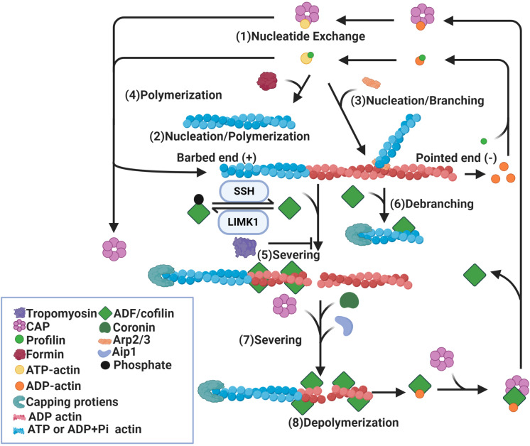
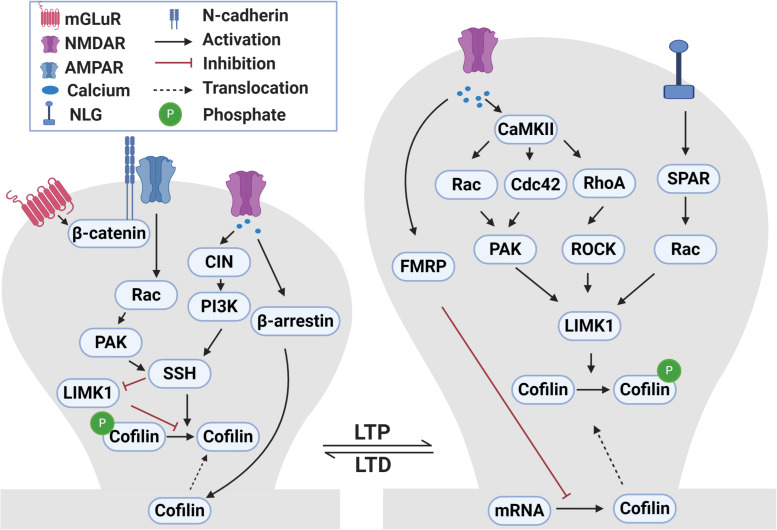
Actin-depolymerization factor (ADF)/cofilin, a family of actin-binding proteins, are critical for the regulation of actin reorganization in response to various signals. Accumulating evidence indicates that ADF/cofilin also play important roles in neuronal structure and function, including long-term potentiation and depression. These are the most extensively studied forms of long-lasting synaptic plasticity and are widely regarded as cellular mechanisms underlying learning and memory. ADF/cofilin regulate synaptic function through their effects on dendritic spines and the trafficking of glutamate receptors, the principal mediator of excitatory synaptic transmission in vertebrates. Regulation of ADF/cofilin involves various signaling pathways converging on LIM domain kinases and slingshot phosphatases, which phosphorylate/inactivate and dephosphorylate/activate ADF/cofilin, respectively. Actin-depolymerization factor/cofilin activity is also regulated by other actin-binding proteins, activity-dependent subcellular distribution and protein translation. Abnormalities in ADF/cofilin have been associated with several neurodegenerative disorders such as Alzheimer's disease. Therefore, investigating the roles of ADF/cofilin in the brain is not only important for understanding the fundamental processes governing neuronal structure and function, but also may provide potential therapeutic strategies to treat brain disorders.

Keywords: ADF/cofilin; AMPA glutamate receptor; LTD; LTP; dendritic spine.


Figures

FIGURE 1
FIGURE 1
Regulation of actin dynamics by ADF/cofilin. Binding of profilin on ADP-actin monomers induces nucleotide exchange (1). Formin (2) and Arp2/3 (3) induce nucleation of actin monomers and formation of actin filaments. While formin induces parallel actin filament (2), Arp2/3 promotes branching of the original filament (3). In addition, actin filaments can be polymerized by the addition of ATP-actin monomers at the barbed ends (4). Binding of dephosphorylated/active ADF/cofilin to ADP-actin subunits of actin filaments causes severing of these filaments (5) and depolymerization at pointed ends (8). ADF/cofilin activity is mediated by phosphorylation and dephosphorylation by LIMK1 and SSH respectively. ADF/cofilin also debranch Arp2/3 nucleated actin filaments (6). The severing activity of ADF/cofilin is enhanced by Aip1, coronin and CAP (7) and diminished by tropomyosin (5). Binding of capping proteins at barbed ends blocks the growth of newly formed actin segments. CAP also dissociates ADF/cofilin from ADP-actin monomers and promotes nucleotide exchange on these monomers (1).
FIGURE 2
FIGURE 2
Signaling pathways that regulate ADF/cofilin phosphorylation and dephosphorylation during LTP and LTD. During LTP, activation of NMDA receptors causes calcium influx into dendritic spines. The increased intracellular calcium activates CaMKII which in turn activates small Rho GTPases, including Rac, Cdc42 and RhoA. These small GTPases bind to and activate PAKs and ROCKs that can directly phosphorylate and activate LIMK1. LIMK1 can also be activated following the activation of neuroligin 1 receptors during LTP through SPAR-Rac signaling pathway. Activated LIMK1 phosphorylates and inactivates cofilin resulting in the enlargement of dendritic spines. In addition, the LTP-induced calcium influx diminishes local translation of cofilin mRNA in dendrites through a FRMP 1-dependent manner. Translocation of cofilin into spines during LTP occurs through yet to be discovered mechanisms. During LTD, the activation of NMDA receptors and influx of calcium activates CIN which activates SSH through the PI3K-dependent pathway. Activated SSH dephosphorylates and activates cofilin which results in dendritic spine shrinkage. In addition, LTD-induced calcium influx mediates translocation of cofilin into spines in a β-arrestin 2-dependent manner. During mGLuR-LTD, GluA2 interaction with cadherin/β-catenin activates Rac-PAK which then activate SSH. Additionally, SSH can also dephosphorylate and inactivate LIMK1.

References

    1. Adolph O., Köster S., Räth M., Georgieff M., Weigt H. U., Engele J., et al. (2007). Rapid increase of glial glutamate uptake via blockade of the protein kinase A pathway. Glia 55 1699–1707. 10.1002/glia.20583 - DOI - PubMed
    1. Agrawal P. B., Joshi M., Savic T., Chen Z., Beggs A. H. (2012). Normal myofibrillar development followed by progressive sarcomeric disruption with actin accumulations in a mouse Cfl2 knockout demonstrates requirement of cofilin-2 for muscle maintenance. Hum. Mol. Genet. 21 2341–2356. 10.1093/hmg/dds053 - DOI - PMC - PubMed
    1. Alhadidi Q., Bin Sayeed M. S., Shah Z. A. (2016). Cofilin as a promising therapeutic target for ischemic and hemorrhagic stroke. Transl. Stroke Res. 7 33–41. 10.1007/s12975-015-0438-2 - DOI - PubMed
    1. Allison D. W., Gelfand V. I., Spector I., Craig A. M. (1998). Role of actin in anchoring postsynaptic receptors in cultured hippocampal neurons: differential attachment of NMDA versus AMPA receptors. J. Neurosci. 18 2423–2436. 10.1523/JNEUROSCI.18-07-02423.1998 - DOI - PMC - PubMed
    1. Alvarez V. A., Sabatini B. L. (2007). Anatomical and physiological plasticity of dendritic spines. Ann. Rev. Neurosci. 30 79–97. 10.1146/annurev.neuro.30.051606.094222 - DOI - PubMed