. 2020 Oct 12:5:181.
doi: 10.12688/wellcomeopenres.16002.2. eCollection 2020.

SARS-CoV-2 RNA detected in blood products from patients with COVID-19 is not associated with infectious virus

Affiliations

SARS-CoV-2 RNA detected in blood products from patients with COVID-19 is not associated with infectious virus

Monique I Andersson et al. Wellcome Open Res. .

Abstract

Background: Laboratory diagnosis of SARS-CoV-2 infection (the cause of COVID-19) uses PCR to detect viral RNA (vRNA) in respiratory samples. SARS-CoV-2 RNA has also been detected in other sample types, but there is limited understanding of the clinical or laboratory significance of its detection in blood. Methods: We undertook a systematic literature review to assimilate the evidence for the frequency of vRNA in blood, and to identify associated clinical characteristics. We performed RT-PCR in serum samples from a UK clinical cohort of acute and convalescent COVID-19 cases (n=212), together with convalescent plasma samples collected by NHS Blood and Transplant (NHSBT) (n=462 additional samples). To determine whether PCR-positive blood samples could pose an infection risk, we attempted virus isolation from a subset of RNA-positive samples. Results: We identified 28 relevant studies, reporting SARS-CoV-2 RNA in 0-76% of blood samples; pooled estimate 10% (95%CI 5-18%). Among serum samples from our clinical cohort, 27/212 (12.7%) had SARS-CoV-2 RNA detected by RT-PCR. RNA detection occurred in samples up to day 20 post symptom onset, and was associated with more severe disease (multivariable odds ratio 7.5). Across all samples collected ≥28 days post symptom onset, 0/494 (0%, 95%CI 0-0.7%) had vRNA detected. Among our PCR-positive samples, cycle threshold (ct) values were high (range 33.5-44.8), suggesting low vRNA copy numbers. PCR-positive sera inoculated into cell culture did not produce any cytopathic effect or yield an increase in detectable SARS-CoV-2 RNA. There was a relationship between RT-PCR negativity and the presence of total SARS-CoV-2 antibody (p=0.02). Conclusions: vRNA was detectable at low viral loads in a minority of serum samples collected in acute infection, but was not associated with infectious SARS-CoV-2 (within the limitations of the assays used). This work helps to inform biosafety precautions for handling blood products from patients with current or previous COVID-19.

Keywords: COVID-19; RNA; SARS-CoV-2; biomarker; blood; laboratory safety; viraemia; viral load.


Conflict of interest statement

Competing interests: DWE has received personal fees from Gilead, outside the submitted work.

Figures
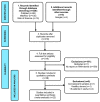
Figure 1.
Figure 1.. PRISMA flow diagram showing number of abstracts identified through a systematic literature review, rejections (with reasons), and final number of studies included in the analysis.
Figure 2.
Figure 2.. Prevalence of SARS-CoV-2 RNA in serum / plasma / whole blood samples from a systematic literature review.
Point prevalence indicated for each study with confidence intervals showing citation and number of samples represented ( Table 1).
Figure 3.
Figure 3.. Relationship between RNA-aemia and days from COVID-19 symptom onset.
Data shown for 212 samples collected from acute and convalescent adults from the Oxford University Hospitals cohort. Positive and negative results are shown plotted at 1 and 0 on the y-axis respectively, with jitter applied to show all points. The line shows the univariable predicted probability of RNA detection over time (95% CI: shaded).
Figure 4.
Figure 4.. Typical images from cell culture in an in vitro system for SARS-CoV-2 culture.
Top row shows controls: ( A) Negative control Vero E6 cells in media; ( B) Cytopathic effect (CPE) in Vero E6 cells spiked with Victoria/01/2020 SARS-CoV-2; Bottom row shows Vero E6 cells inoculated with 1/10 dilution of serum sample from sample VC12 (patient ID UKCOV040), that tested positive for SARS-CoV-2 RNA by RT-PCR; ( C) Aberrant cellular effects at day 4 in a culture inoculated with VC12 at day 0; ( D) Normal appearance of cells at day 7 inoculated with 1/10 dilution of the culture supernatant of the VC12-challenged culture, illustrated in ( C). Raw unedited microscope images can be accessed individually on line .
Figure 5.
Figure 5.. Relationship between serum SARS-COV-2 RT-PCR and total antibody titre, determined by Siemens SARS-CoV-2 Atellica assay.
( A) Proportion of samples testing antibody positive according to RT-PCR status of serum sample; p-value by Fisher’s Exact Test; ( B) Distribution of IgG titres in samples according to RT-PCR status of serum sample; boxes show median/IQR and whiskers show range; p-value by Mann-Whitney U test; ( C) Antibody titres in 23 serum samples testing RT-PCR positive for SARS-CoV-2 RNA, presented in rank order. The bars marked with an asterisk indicate the samples for which in vitro culture was attempted. In all panels, antibody positivity is reported according to the threshold set by the assay manufacturer.

References

    1. World Health Organisation Coronavirus disease (COVID-19) Situation Dashboard. who.int. [cited 31 Mar 2020]. Reference Source
    1. Tang YW, Schmitz JE, Persing DH, et al. : The Laboratory Diagnosis of COVID-19 Infection: Current Issues and Challenges. J Clin Microbiol. 2020;58(6):e00512–20. 10.1128/JCM.00512-20 - DOI - PMC - PubMed
    1. Zheng S, Fan J, Yu F, et al. : Viral load dynamics and disease severity in patients infected with SARS-CoV-2 in Zhejiang province, China, January-March 2020: retrospective cohort study. BMJ. 2020;369:m1443. 10.1136/bmj.m1443 - DOI - PMC - PubMed
    1. Wang W, Xu Y, Gao R, et al. : Detection of SARS-CoV-2 in Different Types of Clinical Specimens. JAMA. 2020;323(18):1843–1844. 10.1001/jama.2020.3786 - DOI - PMC - PubMed
    1. Cheng MP, Papenburg J, Desjardins M, et al. : Diagnostic Testing for Severe Acute Respiratory Syndrome-Related Coronavirus-2: A Narrative Review. Ann Intern Med. 2020;172(11):726–734. 10.7326/M20-1301 - DOI - PMC - PubMed