

Interpretation of Brain Morphology in Association to Alzheimer's Disease Dementia Classification Using Graph Convolutional Networks on Triangulated Meshes
- PMID: 33283214
- PMCID: PMC7713521
- DOI: 10.1007/978-3-030-61056-2_8
Interpretation of Brain Morphology in Association to Alzheimer's Disease Dementia Classification Using Graph Convolutional Networks on Triangulated Meshes
Abstract
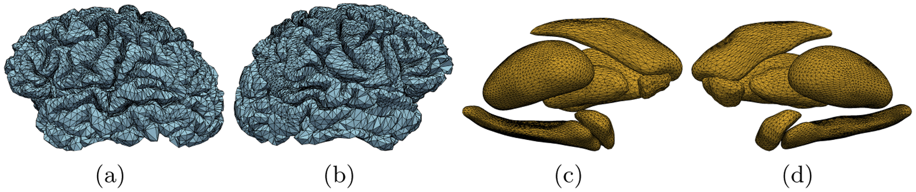
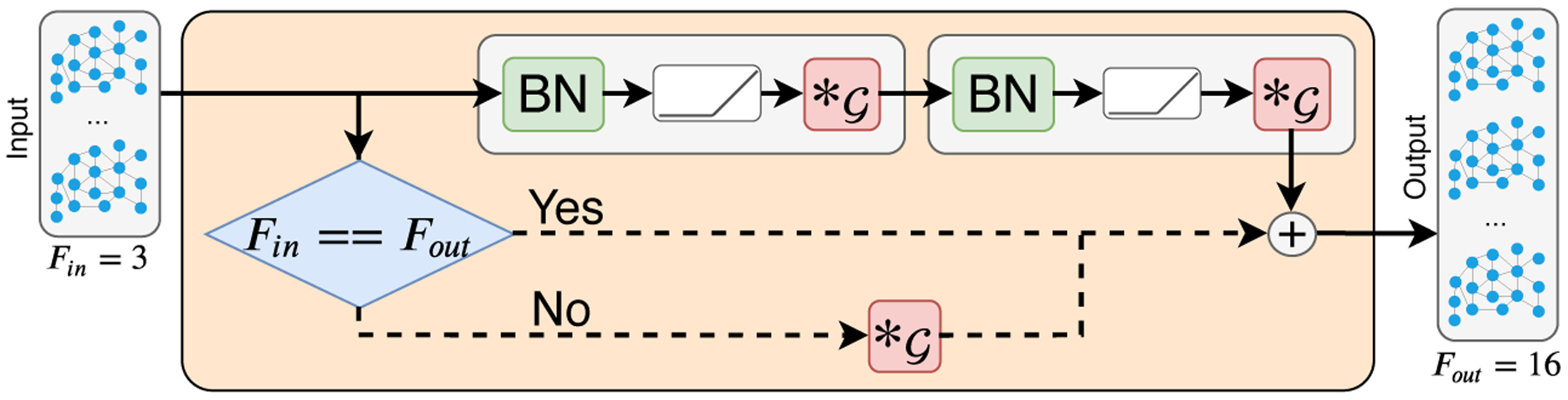
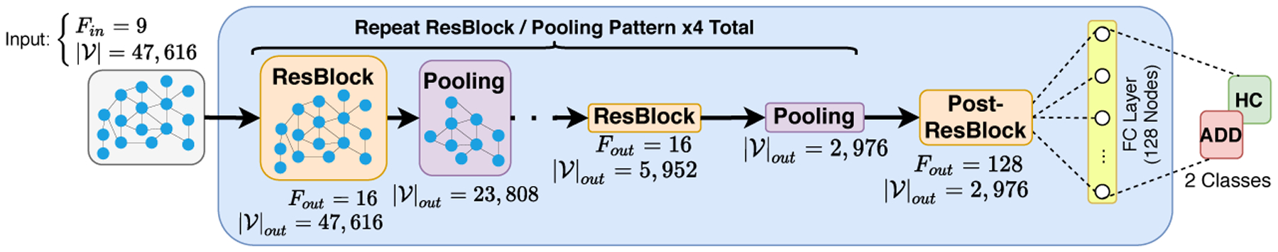
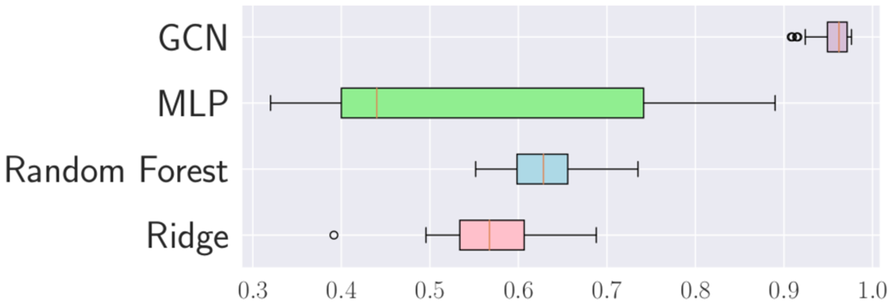
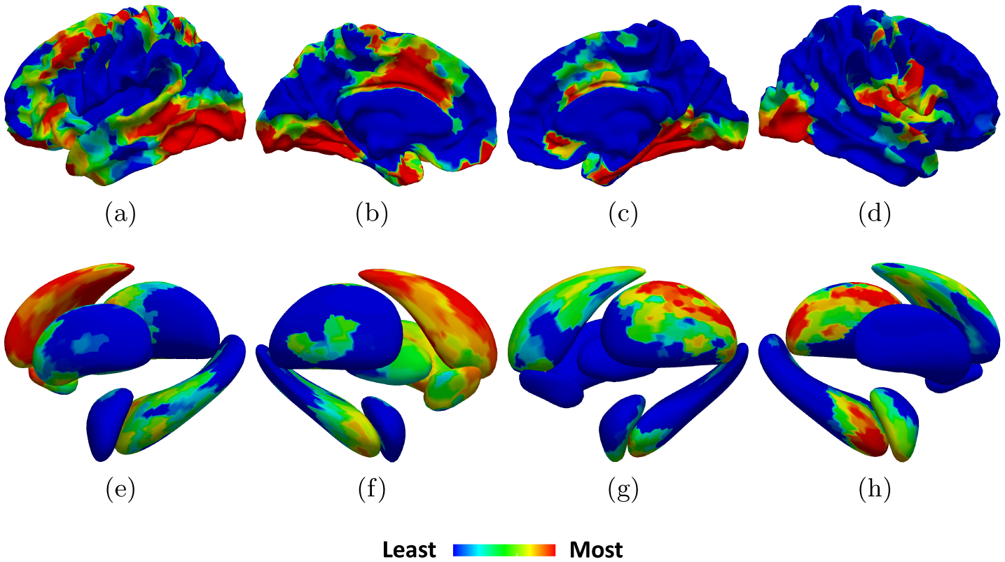
We propose a mesh-based technique to aid in the classification of Alzheimer's disease dementia (ADD) using mesh representations of the cortex and subcortical structures. Deep learning methods for classification tasks that utilize structural neuroimaging often require extensive learning parameters to optimize. Frequently, these approaches for automated medical diagnosis also lack visual interpretability for areas in the brain involved in making a diagnosis. This work: (a) analyzes brain shape using surface information of the cortex and subcortical structures, (b) proposes a residual learning framework for state-of-the-art graph convolutional networks which offer a significant reduction in learnable parameters, and (c) offers visual interpretability of the network via class-specific gradient information that localizes important regions of interest in our inputs. With our proposed method leveraging the use of cortical and subcortical surface information, we outperform other machine learning methods with a 96.35% testing accuracy for the ADD vs. healthy control problem. We confirm the validity of our model by observing its performance in a 25-trial Monte Carlo cross-validation. The generated visualization maps in our study show correspondences with current knowledge regarding the structural localization of pathological changes in the brain associated to dementia of the Alzheimer's type.
Keywords: Alzheimer’s disease classification; Graph convolutional networks; neural network interpretability; triangulated meshes.
Figures
References
-
- Arfken GB, Weber HJ, Harris FE: Mathematical Methods for Physicists. Academic Press, third edn (2013). 10.1016/C2013-0-10310-8 - DOI
-
- Beheshti I, et al. : Classification of alzheimer’s disease and prediction of mild cognitive impairment-to-alzheimer’s conversion from structural magnetic resource imaging using feature ranking and a genetic algorithm. Computers in biology and medicine 83, 109–119 (2017). 10.1016/j.compbiomed.2017.02.011 - DOI - PubMed
-
- Bruyn G: Atlas of the cerebral sulci, vol. 93 G. Thieme Verlag; (1991)
Grants and funding
LinkOut - more resources
Full Text Sources