. 2021 Nov 2;73(9):e3536-e3544.
doi: 10.1093/cid/ciaa1823.

Optic Nerve Sheath Ultrasound for the Detection and Monitoring of Raised Intracranial Pressure in Tuberculous Meningitis

Affiliations

Optic Nerve Sheath Ultrasound for the Detection and Monitoring of Raised Intracranial Pressure in Tuberculous Meningitis

Joseph Donovan et al. Clin Infect Dis. .

Abstract

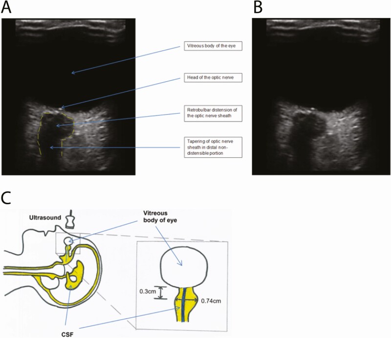
Background: Neurological complications of tuberculous meningitis (TBM) often lead to raised intracranial pressure (ICP) resulting in high morbidity and mortality. Measurement of optic nerve sheath diameter (ONSD) by point-of-care ultrasound may aid in the identification of raised ICP in TBM.

Methods: From June 2017 to December 2019, 107 Vietnamese adults with TBM, enrolled in the ACT HIV or LAST ACT trials (NCT03092817, NCT03100786), underwent ONSD ultrasound at ≥1 of days 0, 3, 7, 14, 21, and day ±30 after enrollment. Demographic data, TBM severity grade, HIV coinfection status, and clinical endpoints by 3 months were recorded. ONSD values were correlated with disease severity, baseline brain imaging, cerebrospinal fluid parameters, and clinical endpoints.

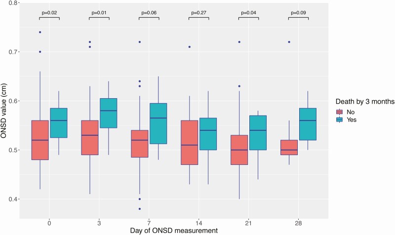
Results: 267 ONSD ultrasound scans were performed in 107 participants over the first 30 days of treatment, with measurements from 0.38-0.74 cm. Paired baseline ONSD and brain imaging were performed in 63 participants. Higher baseline ONSD was associated with more severe disease and abnormal brain imaging (abnormal imaging 0.55 cm vs 0.50 cm normal imaging, P = .01). Baseline median ONSD was significantly higher in participants who died by 3 months (0.56 cm [15/72]) versus participants who survived by 3 months (0.52 cm [57/72]) (P = .02). Median ONSD was higher at all follow-up times in participants who died by 3 months.

Conclusions: Higher ONSD was associated with increased disease severity, brain imaging abnormalities, and increased death by 3 months. ONSD ultrasound has a potential role as a noninvasive, affordable bedside tool for predicting brain pathology and death in TBM.

Keywords: intracranial pressure; optic nerve sheath; tuberculous meningitis; ultrasound.


Figures

Figure 1.
Figure 1.
Distended optic nerve sheath consistent with raised intracranial pressure. Ultrasound images of the right eye are shown with (A) and without (B) descriptive labels. The borders of the optic nerve sheath are marked with a dotted line in panel A. In panel C, a diagram illustrates the appearances seen under ultrasound, showing how these appearances relate to patient position, CSF spaces (yellow) and optic nerve (blue). ONSD measured 0.3 cm from the posterior border of the globe of the eye was 0.74 cm. ONSD is measured as the distance inside the dura mater. Abbreviations: CSF, cerebrospinal fluid; ONSD, optic nerve sheath diameter.
Figure 2.
Figure 2.
ONSD over 30 days of anti-TB chemotherapy, stratified by death by 3 months. For each individual boxplot, the central horizontal bar represents the median value. The box contains data between the third quartile (upper end of box) and the first quartile (lower end of box). Vertical lines above and below each box extend to the most extreme data point that is within 1.5× the vertical height of the box. Dots represent individual data points outside of these limits. P values represent statistical comparison of ONSD values performed by the Wilcoxon rank-sum test. Abbreviations: ONSD, optic nerve sheath diameter; TB, tuberculosis.
Figure 3.
Figure 3.
ONSD over 30 days of anti-TB chemotherapy, stratified by neurological complications by 3 months. For each individual boxplot, the central horizontal bar represents the median value. The box contains data between the third quartile (upper end of box) and the first quartile (lower end of box). Vertical lines above and below each box extend to the most extreme data point that is within 1.5× the vertical height of the box. Dots represent individual data points outside of these limits. P values represent statistical comparison of ONSD values performed by the Wilcoxon rank sum test. Abbreviations: ONSD, optic nerve sheath diameter; TB, tuberculosis.
Figure 4.
Figure 4.
ONSD values over 30 days of anti-TB chemotherapy, stratified by TBM severity grade and death by 3 months. Individual data points represent individual ONSD values at a specified day of measurement. Data are shown stratified by grades 1, 2, and 3, where grade refers to MRC TBM severity grade. Blue dots represent ONSD values in patients who were alive by 3 months, whereas red dots represent ONSD values in patients who died by 3 months. The mean ONSD value across time points, for each grade, is represented by a green line with an associated 95% CI. Abbreviations: CI, confidence interval; MRC, Medical Research Council; ONSD, optic nerve sheath diameter; TB, tuberculosis; TBM, tuberculous meningitis.

References

    1. Christensen AS, Roed C, Omland LH, Andersen PH, Obel N, Andersen ÅB. Long-term mortality in patients with tuberculous meningitis: a Danish nationwide cohort study. PLoS One 2011; 6:e27900. - PMC - PubMed
    1. Vinnard C, King L, Munsiff S, et al. Long-term mortality of patients with tuberculous meningitis in New York city: a cohort study. Clin Infect Dis 2016; 64:ciw763. - PMC - PubMed
    1. Thwaites GE, Nguyen DB, Nguyen HD, et al. Dexamethasone for the treatment of tuberculous meningitis in adolescents and adults. N Engl J Med 2004; 351:1741–51. - PubMed
    1. Ruslami R, Ganiem AR, Dian S, et al. Intensified regimen containing rifampicin and moxifloxacin for tuberculous meningitis: an open-label, randomised controlled phase 2 trial. Lancet Infect Dis 2013; 13:27–35. - PubMed
    1. Donovan J, Figaji A, Imran D, Phu NH, Rohlwink U, Thwaites GE. The neurocritical care of tuberculous meningitis. Lancet Neurol 2019; 18:771–83. - PubMed

Publication types

Associated data