Are sleep paralysis and false awakenings different from REM sleep and from lucid REM sleep? A spectral EEG analysis
- PMID: 33283752
- PMCID: PMC8020694
- DOI: 10.5664/jcsm.9056
Are sleep paralysis and false awakenings different from REM sleep and from lucid REM sleep? A spectral EEG analysis
Abstract
Study objectives: To determine the polysomnography characteristics during sleep paralysis, false awakenings, and lucid dreaming (which are states intermediate to rapid eye movement [REM] sleep and wake but exceptionally observed in sleep laboratory).
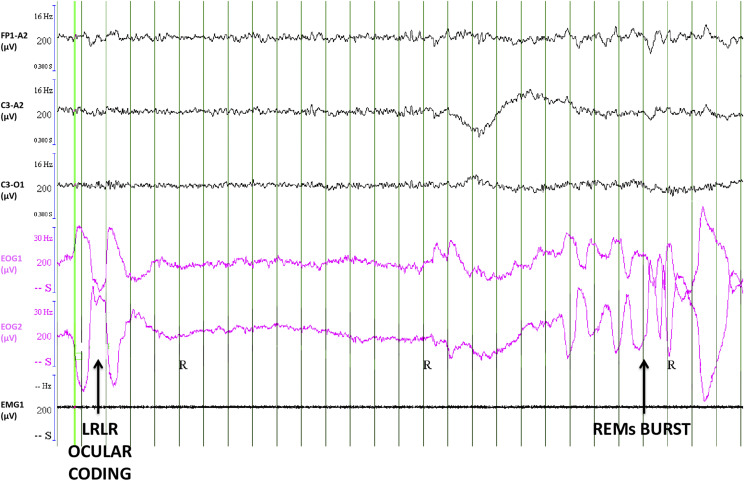
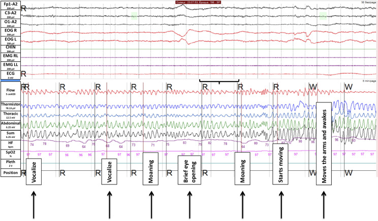
Methods: In 5 participants, we captured 5 episodes of sleep paralysis (2 time marked with the ocular left-right-left-right code normally used to signal lucid dreaming, 1 time marked by an external noise, and 2 retrospectively reported) and 2 episodes of false awakening. The sleep coding (using 3-second mini-epochs) and spectral electroencephalography analysis were compared during these episodes and normal REM sleep as well as wakefulness in the same 4 of 5 participants and vs lucid REM sleep in 4 other patients with narcolepsy.
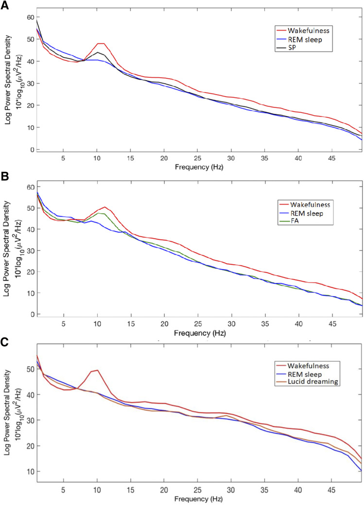
Results: During episodes of sleep paralysis, 70.8% of mini-epochs contained theta electroencephalography rhythm (vs 89.7% in REM sleep and 21.2% in wakefulness), 93.8% contained chin muscle atonia (vs 89.7% in REM sleep and 33.3% in wakefulness), and 6.9% contained rapid eye movements (vs 11.9% in REM sleep and 8.1% in wakefulness). The electroencephalography spectrum during sleep paralysis was intermediate between wakefulness and REM sleep in the alpha, theta, and delta frequencies, whereas the beta frequencies were not different between sleep paralysis and normal REM sleep. The power spectrum during false awakening followed the same profile as in sleep paralysis.
Conclusions: The predominant theta electroencephalography rhythm during sleep paralysis and false awakenings (with rare and lower alpha rhythm) suggests that the brain during sleep paralysis is not in an awake but in a dreaming state.
Keywords: REM sleep; false awakenings; sleep paralysis.
© 2021 American Academy of Sleep Medicine.
Conflict of interest statement
All authors have seen and approved this manuscript. The authors report no conflicts of interest.
Figures
References
-
- Fox K, Christoff K. The Oxford Handbook of Spontaneous Thought: Mind-Wandering, Creativity, and Dreaming. Oxford University Press: Oxford, UK; 2018.
-
- American Academy of Sleep Medicine . International Classification of Sleep Disorders. 3rd ed. Darien, IL:American Academy of Sleep Medicine; 2014.
MeSH terms
LinkOut - more resources
Full Text Sources
Other Literature Sources